Magnesium »
PDB 1qc5-1qsh »
1qm4 »
Magnesium in PDB 1qm4: Methionine Adenosyltransferase Complexed with A L-Methionine Analogue
Enzymatic activity of Methionine Adenosyltransferase Complexed with A L-Methionine Analogue
All present enzymatic activity of Methionine Adenosyltransferase Complexed with A L-Methionine Analogue:
2.5.1.6;
2.5.1.6;
Protein crystallography data
The structure of Methionine Adenosyltransferase Complexed with A L-Methionine Analogue, PDB code: 1qm4
was solved by
B.Gonzalez,
M.A.Pajares,
J.A.Hermoso,
J.Sanz-Aparicio,
with X-Ray Crystallography technique. A brief refinement statistics is given in the table below:
| Resolution Low / High (Å) | 10.00 / 2.66 |
| Space group | P 41 2 2 |
| Cell size a, b, c (Å), α, β, γ (°) | 115.200, 115.200, 159.980, 90.00, 90.00, 90.00 |
| R / Rfree (%) | 23 / 29 |
Other elements in 1qm4:
The structure of Methionine Adenosyltransferase Complexed with A L-Methionine Analogue also contains other interesting chemical elements:
| Potassium | (K) | 3 atoms |
Magnesium Binding Sites:
The binding sites of Magnesium atom in the Methionine Adenosyltransferase Complexed with A L-Methionine Analogue
(pdb code 1qm4). This binding sites where shown within
5.0 Angstroms radius around Magnesium atom.
In total 2 binding sites of Magnesium where determined in the Methionine Adenosyltransferase Complexed with A L-Methionine Analogue, PDB code: 1qm4:
Jump to Magnesium binding site number: 1; 2;
In total 2 binding sites of Magnesium where determined in the Methionine Adenosyltransferase Complexed with A L-Methionine Analogue, PDB code: 1qm4:
Jump to Magnesium binding site number: 1; 2;
Magnesium binding site 1 out of 2 in 1qm4
Go back to
Magnesium binding site 1 out
of 2 in the Methionine Adenosyltransferase Complexed with A L-Methionine Analogue
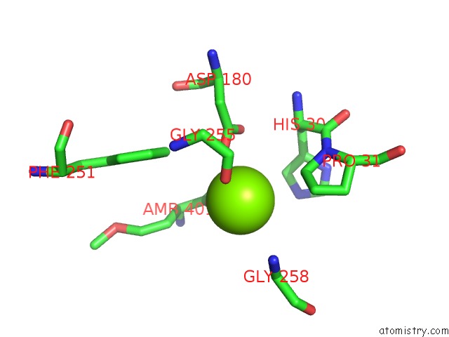
Mono view
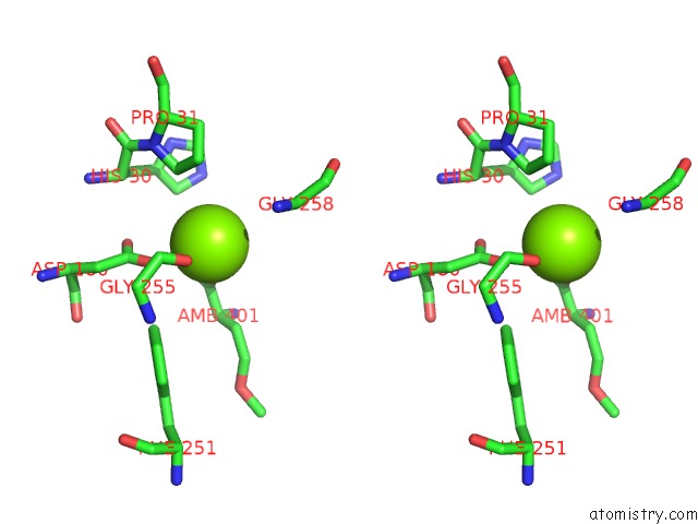
Stereo pair view
Mono view
Stereo pair view
A full contact list of Magnesium with other atoms in the Mg binding
site number 1 of Methionine Adenosyltransferase Complexed with A L-Methionine Analogue within 5.0Å range:
|
Magnesium binding site 2 out of 2 in 1qm4
Go back to
Magnesium binding site 2 out
of 2 in the Methionine Adenosyltransferase Complexed with A L-Methionine Analogue
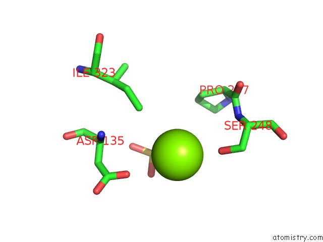
Mono view
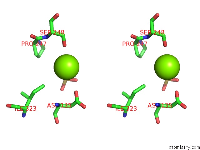
Stereo pair view
Mono view
Stereo pair view
A full contact list of Magnesium with other atoms in the Mg binding
site number 2 of Methionine Adenosyltransferase Complexed with A L-Methionine Analogue within 5.0Å range:
|
Reference:
B.Gonzalez,
M.A.Pajares,
J.A.Hermoso,
L.Alvarez,
F.Garrido,
J.R.Sufrin,
J.Sanz-Aparicio.
The Crystal Structure of Tetrameric Methionine Adenosyltransferase From Rat Liver Reveals the Methionine-Binding Site J.Mol.Biol. V. 300 363 2000.
ISSN: ISSN 0022-2836
PubMed: 10873471
DOI: 10.1006/JMBI.2000.3858
Page generated: Tue Aug 13 11:53:36 2024
ISSN: ISSN 0022-2836
PubMed: 10873471
DOI: 10.1006/JMBI.2000.3858
Last articles
Zn in 9J0NZn in 9J0O
Zn in 9J0P
Zn in 9FJX
Zn in 9EKB
Zn in 9C0F
Zn in 9CAH
Zn in 9CH0
Zn in 9CH3
Zn in 9CH1