Magnesium »
PDB 1qsi-1rc5 »
1r03 »
Magnesium in PDB 1r03: Crystal Structure of A Human Mitochondrial Ferritin
Protein crystallography data
The structure of Crystal Structure of A Human Mitochondrial Ferritin, PDB code: 1r03
was solved by
B.Corsi,
P.Santambrogio,
P.Arosio,
S.Levi,
B.Langlois D'estaintot,
T.Granier,
B.Gallois,
J.M.Chevallier,
G.Precigoux,
with X-Ray Crystallography technique. A brief refinement statistics is given in the table below:
| Resolution Low / High (Å) | 14.90 / 1.70 |
| Space group | F 4 3 2 |
| Cell size a, b, c (Å), α, β, γ (°) | 181.410, 181.410, 181.410, 90.00, 90.00, 90.00 |
| R / Rfree (%) | 17 / 20 |
Magnesium Binding Sites:
The binding sites of Magnesium atom in the Crystal Structure of A Human Mitochondrial Ferritin
(pdb code 1r03). This binding sites where shown within
5.0 Angstroms radius around Magnesium atom.
In total 5 binding sites of Magnesium where determined in the Crystal Structure of A Human Mitochondrial Ferritin, PDB code: 1r03:
Jump to Magnesium binding site number: 1; 2; 3; 4; 5;
In total 5 binding sites of Magnesium where determined in the Crystal Structure of A Human Mitochondrial Ferritin, PDB code: 1r03:
Jump to Magnesium binding site number: 1; 2; 3; 4; 5;
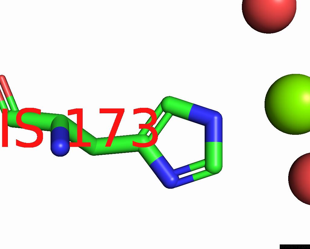
Magnesium binding site 1 out of 5 in 1r03
Go back to
Magnesium binding site 1 out
of 5 in the Crystal Structure of A Human Mitochondrial Ferritin

Mono view

Stereo pair view
Mono view
Stereo pair view
A full contact list of Magnesium with other atoms in the Mg binding
site number 1 of Crystal Structure of A Human Mitochondrial Ferritin within 5.0Å range:
|
Magnesium binding site 2 out of 5 in 1r03
Go back to
Magnesium binding site 2 out
of 5 in the Crystal Structure of A Human Mitochondrial Ferritin
Mono view

Stereo pair view
Mono view
Stereo pair view
A full contact list of Magnesium with other atoms in the Mg binding
site number 2 of Crystal Structure of A Human Mitochondrial Ferritin within 5.0Å range:
|
Magnesium binding site 3 out of 5 in 1r03
Go back to
Magnesium binding site 3 out
of 5 in the Crystal Structure of A Human Mitochondrial Ferritin

Mono view

Stereo pair view
Mono view
Stereo pair view
A full contact list of Magnesium with other atoms in the Mg binding
site number 3 of Crystal Structure of A Human Mitochondrial Ferritin within 5.0Å range:
|
Magnesium binding site 4 out of 5 in 1r03
Go back to
Magnesium binding site 4 out
of 5 in the Crystal Structure of A Human Mitochondrial Ferritin

Mono view

Stereo pair view
Mono view
Stereo pair view
A full contact list of Magnesium with other atoms in the Mg binding
site number 4 of Crystal Structure of A Human Mitochondrial Ferritin within 5.0Å range:
|
Magnesium binding site 5 out of 5 in 1r03
Go back to
Magnesium binding site 5 out
of 5 in the Crystal Structure of A Human Mitochondrial Ferritin

Mono view

Stereo pair view
Mono view
Stereo pair view
A full contact list of Magnesium with other atoms in the Mg binding
site number 5 of Crystal Structure of A Human Mitochondrial Ferritin within 5.0Å range:
|
Reference:
B.Langlois D'estaintot,
P.Santambrogio,
T.Granier,
B.Gallois,
J.M.Chevallier,
G.Precigoux,
S.Levi,
P.Arosio.
Crystal Structure and Biochemical Properties of the Human Mitochondrial Ferritin and Its Mutant SER144ALA J.Mol.Biol. V. 340 277 2004.
ISSN: ISSN 0022-2836
PubMed: 15201052
DOI: 10.1016/J.JMB.2004.04.036
Page generated: Tue Aug 13 11:58:02 2024
ISSN: ISSN 0022-2836
PubMed: 15201052
DOI: 10.1016/J.JMB.2004.04.036
Last articles
Ca in 5TYUCa in 5TYC
Ca in 5TWT
Ca in 5TYB
Ca in 5TXZ
Ca in 5TXX
Ca in 5TX6
Ca in 5TVQ
Ca in 5TUA
Ca in 5TSP