Magnesium »
PDB 1qsi-1rc5 »
1r2q »
Magnesium in PDB 1r2q: Crystal Structure of Human RAB5A Gtpase Domain at 1.05 A Resolution
Protein crystallography data
The structure of Crystal Structure of Human RAB5A Gtpase Domain at 1.05 A Resolution, PDB code: 1r2q
was solved by
S.Terzyan,
G.Zhu,
G.Li,
X.C.Zhang,
with X-Ray Crystallography technique. A brief refinement statistics is given in the table below:
| Resolution Low / High (Å) | 29.02 / 1.05 |
| Space group | P 21 21 21 |
| Cell size a, b, c (Å), α, β, γ (°) | 35.727, 64.573, 66.180, 90.00, 90.00, 90.00 |
| R / Rfree (%) | 12.4 / 16.9 |
Magnesium Binding Sites:
The binding sites of Magnesium atom in the Crystal Structure of Human RAB5A Gtpase Domain at 1.05 A Resolution
(pdb code 1r2q). This binding sites where shown within
5.0 Angstroms radius around Magnesium atom.
In total only one binding site of Magnesium was determined in the Crystal Structure of Human RAB5A Gtpase Domain at 1.05 A Resolution, PDB code: 1r2q:
In total only one binding site of Magnesium was determined in the Crystal Structure of Human RAB5A Gtpase Domain at 1.05 A Resolution, PDB code: 1r2q:
Magnesium binding site 1 out of 1 in 1r2q
Go back to
Magnesium binding site 1 out
of 1 in the Crystal Structure of Human RAB5A Gtpase Domain at 1.05 A Resolution
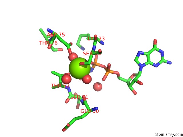
Mono view
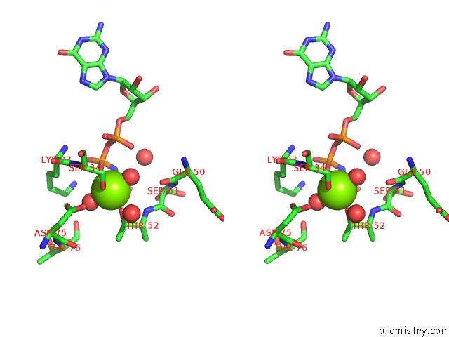
Stereo pair view
Mono view
Stereo pair view
A full contact list of Magnesium with other atoms in the Mg binding
site number 1 of Crystal Structure of Human RAB5A Gtpase Domain at 1.05 A Resolution within 5.0Å range:
|
Reference:
S.Terzyan,
G.Zhu,
G.Li,
X.C.Zhang.
Refinement of the Structure of Human RAB5A Gtpase Domain at 1.05 A Resolution. Acta Crystallogr.,Sect.D V. 60 54 2004.
ISSN: ISSN 0907-4449
PubMed: 14684892
DOI: 10.1107/S0907444903021632
Page generated: Tue Aug 13 12:01:27 2024
ISSN: ISSN 0907-4449
PubMed: 14684892
DOI: 10.1107/S0907444903021632
Last articles
Zn in 9J0NZn in 9J0O
Zn in 9J0P
Zn in 9FJX
Zn in 9EKB
Zn in 9C0F
Zn in 9CAH
Zn in 9CH0
Zn in 9CH3
Zn in 9CH1