Magnesium »
PDB 1qsi-1rc5 »
1r3c »
Magnesium in PDB 1r3c: The Structure of P38ALPHA C162S Mutant
Enzymatic activity of The Structure of P38ALPHA C162S Mutant
All present enzymatic activity of The Structure of P38ALPHA C162S Mutant:
2.7.1.37;
2.7.1.37;
Protein crystallography data
The structure of The Structure of P38ALPHA C162S Mutant, PDB code: 1r3c
was solved by
S.B.Patel,
P.M.Cameron,
B.Frantz-Wattley,
E.O'neill,
J.W.Becker,
G.Scapin,
with X-Ray Crystallography technique. A brief refinement statistics is given in the table below:
| Resolution Low / High (Å) | 17.00 / 2.00 |
| Space group | P 21 21 21 |
| Cell size a, b, c (Å), α, β, γ (°) | 45.162, 84.799, 124.908, 90.00, 90.00, 90.00 |
| R / Rfree (%) | 20 / 22.4 |
Magnesium Binding Sites:
The binding sites of Magnesium atom in the The Structure of P38ALPHA C162S Mutant
(pdb code 1r3c). This binding sites where shown within
5.0 Angstroms radius around Magnesium atom.
In total 2 binding sites of Magnesium where determined in the The Structure of P38ALPHA C162S Mutant, PDB code: 1r3c:
Jump to Magnesium binding site number: 1; 2;
In total 2 binding sites of Magnesium where determined in the The Structure of P38ALPHA C162S Mutant, PDB code: 1r3c:
Jump to Magnesium binding site number: 1; 2;
Magnesium binding site 1 out of 2 in 1r3c
Go back to
Magnesium binding site 1 out
of 2 in the The Structure of P38ALPHA C162S Mutant
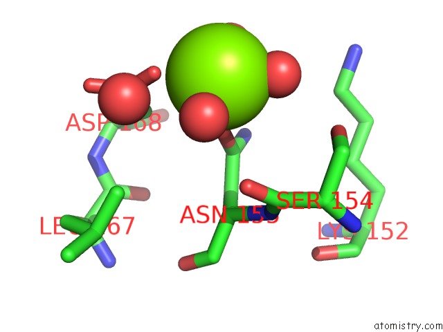
Mono view
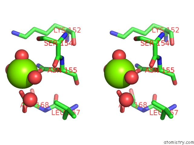
Stereo pair view
Mono view
Stereo pair view
A full contact list of Magnesium with other atoms in the Mg binding
site number 1 of The Structure of P38ALPHA C162S Mutant within 5.0Å range:
|
Magnesium binding site 2 out of 2 in 1r3c
Go back to
Magnesium binding site 2 out
of 2 in the The Structure of P38ALPHA C162S Mutant

Mono view
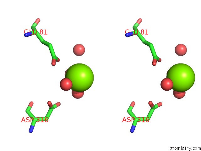
Stereo pair view
Mono view
Stereo pair view
A full contact list of Magnesium with other atoms in the Mg binding
site number 2 of The Structure of P38ALPHA C162S Mutant within 5.0Å range:
|
Reference:
S.B.Patel,
P.M.Cameron,
B.Frantz-Wattley,
E.O'neill,
J.W.Becker,
G.Scapin.
Lattice Stabilization and Enhanced Diffraction in Human P38 Alpha Crystals By Protein Engineering. Biochim.Biophys.Acta V.1696 67 2004.
ISSN: ISSN 0006-3002
PubMed: 14726206
DOI: 10.1016/J.BBAPAP.2003.09.009
Page generated: Tue Aug 13 12:01:53 2024
ISSN: ISSN 0006-3002
PubMed: 14726206
DOI: 10.1016/J.BBAPAP.2003.09.009
Last articles
Zn in 9MJ5Zn in 9HNW
Zn in 9G0L
Zn in 9FNE
Zn in 9DZN
Zn in 9E0I
Zn in 9D32
Zn in 9DAK
Zn in 8ZXC
Zn in 8ZUF