Magnesium »
PDB 1rdd-1rtk »
1rep »
Magnesium in PDB 1rep: Crystal Structure of Replication Initiator Protein REPE54 of Mini-F Plasmid Complexed with An Iteron Dna
Protein crystallography data
The structure of Crystal Structure of Replication Initiator Protein REPE54 of Mini-F Plasmid Complexed with An Iteron Dna, PDB code: 1rep
was solved by
H.Komori,
F.Matsunaga,
Y.Higuchi,
M.Ishiai,
C.Wada,
K.Miki,
with X-Ray Crystallography technique. A brief refinement statistics is given in the table below:
| Resolution Low / High (Å) | 6.00 / 2.60 |
| Space group | C 1 2 1 |
| Cell size a, b, c (Å), α, β, γ (°) | 108.435, 81.853, 73.891, 90.00, 121.53, 90.00 |
| R / Rfree (%) | 21.3 / 27.4 |
Magnesium Binding Sites:
The binding sites of Magnesium atom in the Crystal Structure of Replication Initiator Protein REPE54 of Mini-F Plasmid Complexed with An Iteron Dna
(pdb code 1rep). This binding sites where shown within
5.0 Angstroms radius around Magnesium atom.
In total only one binding site of Magnesium was determined in the Crystal Structure of Replication Initiator Protein REPE54 of Mini-F Plasmid Complexed with An Iteron Dna, PDB code: 1rep:
In total only one binding site of Magnesium was determined in the Crystal Structure of Replication Initiator Protein REPE54 of Mini-F Plasmid Complexed with An Iteron Dna, PDB code: 1rep:
Magnesium binding site 1 out of 1 in 1rep
Go back to
Magnesium binding site 1 out
of 1 in the Crystal Structure of Replication Initiator Protein REPE54 of Mini-F Plasmid Complexed with An Iteron Dna
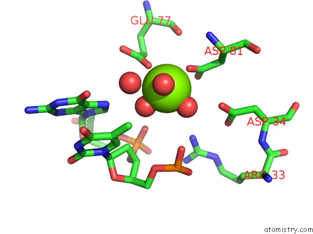
Mono view
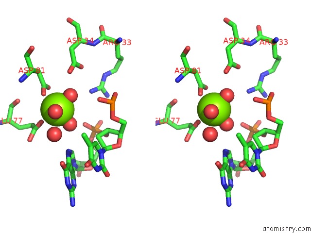
Stereo pair view
Mono view
Stereo pair view
A full contact list of Magnesium with other atoms in the Mg binding
site number 1 of Crystal Structure of Replication Initiator Protein REPE54 of Mini-F Plasmid Complexed with An Iteron Dna within 5.0Å range:
|
Reference:
H.Komori,
F.Matsunaga,
Y.Higuchi,
M.Ishiai,
C.Wada,
K.Miki.
Crystal Structure of A Prokaryotic Replication Initiator Protein Bound to Dna at 2.6 A Resolution. Embo J. V. 18 4597 1999.
ISSN: ISSN 0261-4189
PubMed: 10469640
DOI: 10.1093/EMBOJ/18.17.4597
Page generated: Tue Aug 13 12:59:05 2024
ISSN: ISSN 0261-4189
PubMed: 10469640
DOI: 10.1093/EMBOJ/18.17.4597
Last articles
Zn in 9J0NZn in 9J0O
Zn in 9J0P
Zn in 9FJX
Zn in 9EKB
Zn in 9C0F
Zn in 9CAH
Zn in 9CH0
Zn in 9CH3
Zn in 9CH1