Magnesium »
PDB 1rdd-1rtk »
1rif »
Magnesium in PDB 1rif: Crystal Structure of the Uvsw Helicase From Bacteriophage T4
Protein crystallography data
The structure of Crystal Structure of the Uvsw Helicase From Bacteriophage T4, PDB code: 1rif
was solved by
E.A.Sickmier,
S.W.White,
K.N.Kreuzer,
with X-Ray Crystallography technique. A brief refinement statistics is given in the table below:
| Resolution Low / High (Å) | 20.00 / 2.00 |
| Space group | P 1 21 1 |
| Cell size a, b, c (Å), α, β, γ (°) | 42.739, 95.181, 86.278, 90.00, 95.40, 90.00 |
| R / Rfree (%) | 21.8 / 24.6 |
Other elements in 1rif:
The structure of Crystal Structure of the Uvsw Helicase From Bacteriophage T4 also contains other interesting chemical elements:
| Gold | (Au) | 4 atoms |
Magnesium Binding Sites:
The binding sites of Magnesium atom in the Crystal Structure of the Uvsw Helicase From Bacteriophage T4
(pdb code 1rif). This binding sites where shown within
5.0 Angstroms radius around Magnesium atom.
In total 3 binding sites of Magnesium where determined in the Crystal Structure of the Uvsw Helicase From Bacteriophage T4, PDB code: 1rif:
Jump to Magnesium binding site number: 1; 2; 3;
In total 3 binding sites of Magnesium where determined in the Crystal Structure of the Uvsw Helicase From Bacteriophage T4, PDB code: 1rif:
Jump to Magnesium binding site number: 1; 2; 3;
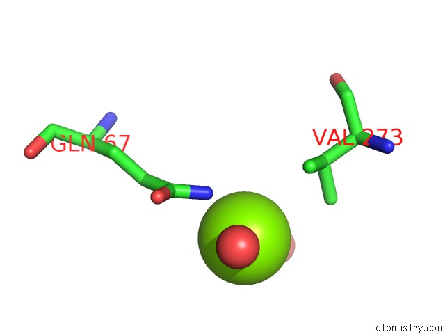
Magnesium binding site 1 out of 3 in 1rif
Go back to
Magnesium binding site 1 out
of 3 in the Crystal Structure of the Uvsw Helicase From Bacteriophage T4
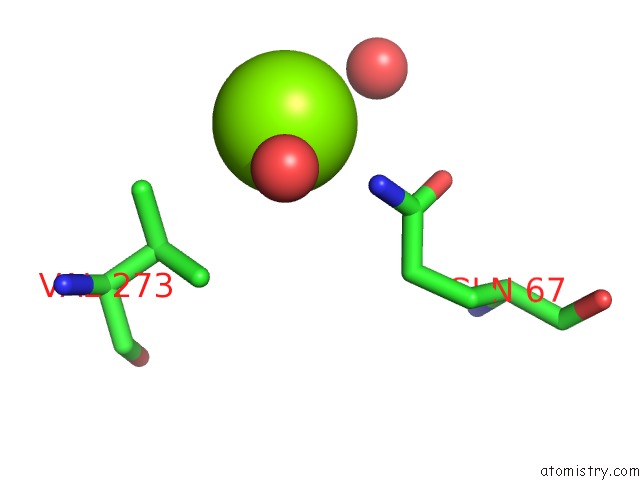
Mono view
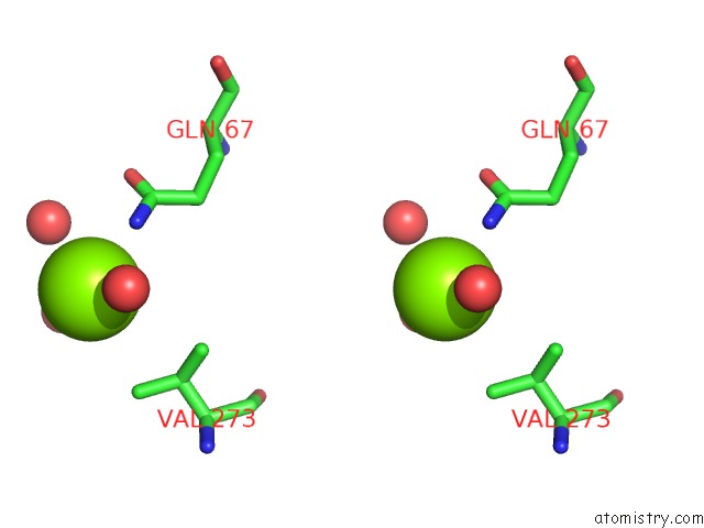
Stereo pair view
Mono view
Stereo pair view
A full contact list of Magnesium with other atoms in the Mg binding
site number 1 of Crystal Structure of the Uvsw Helicase From Bacteriophage T4 within 5.0Å range:
|
Magnesium binding site 2 out of 3 in 1rif
Go back to
Magnesium binding site 2 out
of 3 in the Crystal Structure of the Uvsw Helicase From Bacteriophage T4
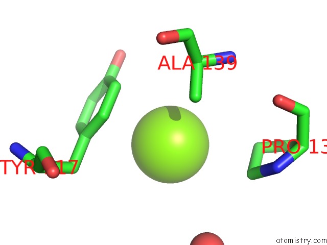
Mono view

Stereo pair view
Mono view
Stereo pair view
A full contact list of Magnesium with other atoms in the Mg binding
site number 2 of Crystal Structure of the Uvsw Helicase From Bacteriophage T4 within 5.0Å range:
|
Magnesium binding site 3 out of 3 in 1rif
Go back to
Magnesium binding site 3 out
of 3 in the Crystal Structure of the Uvsw Helicase From Bacteriophage T4
Mono view

Stereo pair view
Mono view
Stereo pair view
A full contact list of Magnesium with other atoms in the Mg binding
site number 3 of Crystal Structure of the Uvsw Helicase From Bacteriophage T4 within 5.0Å range:
|
Reference:
E.A.Sickmier,
K.N.Kreuzer,
S.W.White.
The Crystal Structure of the Uvsw Helicase From Bacteriophage T4. Structure V. 12 583 2004.
ISSN: ISSN 0969-2126
PubMed: 15062081
DOI: 10.1016/J.STR.2004.02.016
Page generated: Tue Aug 13 13:00:07 2024
ISSN: ISSN 0969-2126
PubMed: 15062081
DOI: 10.1016/J.STR.2004.02.016
Last articles
Cl in 7T1MCl in 7T1L
Cl in 7T1K
Cl in 7T0C
Cl in 7SZE
Cl in 7T0E
Cl in 7SZF
Cl in 7SZQ
Cl in 7SZP
Cl in 7SZH