Magnesium »
PDB 1rtz-1s8f »
1s5j »
Magnesium in PDB 1s5j: Insight in Dna Replication: the Crystal Structure of Dna Polymerase B1 From the Archaeon Sulfolobus Solfataricus
Enzymatic activity of Insight in Dna Replication: the Crystal Structure of Dna Polymerase B1 From the Archaeon Sulfolobus Solfataricus
All present enzymatic activity of Insight in Dna Replication: the Crystal Structure of Dna Polymerase B1 From the Archaeon Sulfolobus Solfataricus:
2.7.7.7;
2.7.7.7;
Protein crystallography data
The structure of Insight in Dna Replication: the Crystal Structure of Dna Polymerase B1 From the Archaeon Sulfolobus Solfataricus, PDB code: 1s5j
was solved by
C.Savino,
L.Federici,
V.Nastopoulos,
K.A.Johnson,
F.M.Pisani,
M.Rossi,
D.Tsernoglou,
with X-Ray Crystallography technique. A brief refinement statistics is given in the table below:
| Resolution Low / High (Å) | 30.00 / 2.40 |
| Space group | C 1 2 1 |
| Cell size a, b, c (Å), α, β, γ (°) | 187.470, 68.800, 125.850, 90.00, 107.94, 90.00 |
| R / Rfree (%) | 23.2 / 27.2 |
Magnesium Binding Sites:
The binding sites of Magnesium atom in the Insight in Dna Replication: the Crystal Structure of Dna Polymerase B1 From the Archaeon Sulfolobus Solfataricus
(pdb code 1s5j). This binding sites where shown within
5.0 Angstroms radius around Magnesium atom.
In total only one binding site of Magnesium was determined in the Insight in Dna Replication: the Crystal Structure of Dna Polymerase B1 From the Archaeon Sulfolobus Solfataricus, PDB code: 1s5j:
In total only one binding site of Magnesium was determined in the Insight in Dna Replication: the Crystal Structure of Dna Polymerase B1 From the Archaeon Sulfolobus Solfataricus, PDB code: 1s5j:
Magnesium binding site 1 out of 1 in 1s5j
Go back to
Magnesium binding site 1 out
of 1 in the Insight in Dna Replication: the Crystal Structure of Dna Polymerase B1 From the Archaeon Sulfolobus Solfataricus
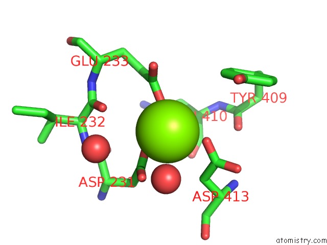
Mono view
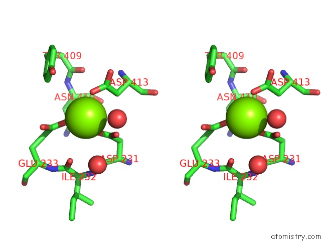
Stereo pair view
Mono view
Stereo pair view
A full contact list of Magnesium with other atoms in the Mg binding
site number 1 of Insight in Dna Replication: the Crystal Structure of Dna Polymerase B1 From the Archaeon Sulfolobus Solfataricus within 5.0Å range:
|
Reference:
C.Savino,
L.Federici,
K.A.Johnson,
B.Vallone,
V.Nastopoulos,
M.Rossi,
F.M.Pisani,
D.Tsernoglou.
Insights Into Dna Replication: the Crystal Structure of Dna Polymerase B1 From the Archaeon Sulfolobus Solfataricus Structure V. 12 2001 2004.
ISSN: ISSN 0969-2126
PubMed: 15530364
DOI: 10.1016/J.STR.2004.09.007
Page generated: Sun Aug 10 04:03:59 2025
ISSN: ISSN 0969-2126
PubMed: 15530364
DOI: 10.1016/J.STR.2004.09.007
Last articles
Mg in 3CC2Mg in 3CCE
Mg in 3CC7
Mg in 3CC4
Mg in 3CB3
Mg in 3CC6
Mg in 3C9U
Mg in 3CBT
Mg in 3CBQ
Mg in 3CBG