Magnesium »
PDB 1s9d-1so5 »
1saw »
Magnesium in PDB 1saw: X-Ray Structure of Homo Sapiens Protein FLJ36880
Protein crystallography data
The structure of X-Ray Structure of Homo Sapiens Protein FLJ36880, PDB code: 1saw
was solved by
B.A.Manjasetty,
F.H.Niesen,
H.Delbrueck,
F.Goetz,
V.Sievert,
K.Buessow,
J.Behlke,
U.Heinemann,
with X-Ray Crystallography technique. A brief refinement statistics is given in the table below:
| Resolution Low / High (Å) | 19.65 / 2.20 |
| Space group | P 21 21 21 |
| Cell size a, b, c (Å), α, β, γ (°) | 50.792, 77.239, 114.195, 90.00, 90.00, 90.00 |
| R / Rfree (%) | 20.4 / 23.9 |
Other elements in 1saw:
The structure of X-Ray Structure of Homo Sapiens Protein FLJ36880 also contains other interesting chemical elements:
| Chlorine | (Cl) | 1 atom |
Magnesium Binding Sites:
The binding sites of Magnesium atom in the X-Ray Structure of Homo Sapiens Protein FLJ36880
(pdb code 1saw). This binding sites where shown within
5.0 Angstroms radius around Magnesium atom.
In total 3 binding sites of Magnesium where determined in the X-Ray Structure of Homo Sapiens Protein FLJ36880, PDB code: 1saw:
Jump to Magnesium binding site number: 1; 2; 3;
In total 3 binding sites of Magnesium where determined in the X-Ray Structure of Homo Sapiens Protein FLJ36880, PDB code: 1saw:
Jump to Magnesium binding site number: 1; 2; 3;
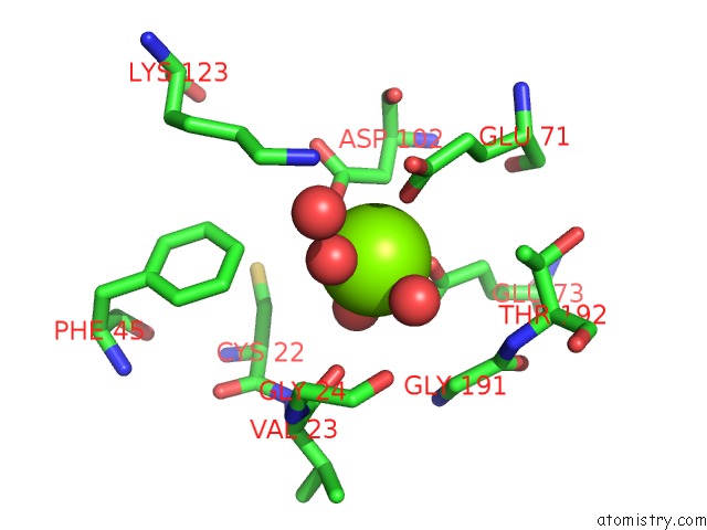
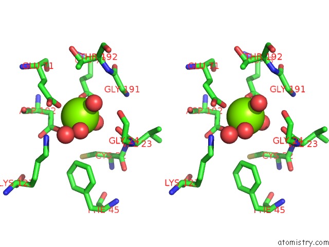
Magnesium binding site 1 out of 3 in 1saw
Go back to
Magnesium binding site 1 out
of 3 in the X-Ray Structure of Homo Sapiens Protein FLJ36880
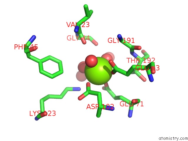
Mono view
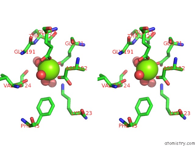
Stereo pair view
Mono view
Stereo pair view
A full contact list of Magnesium with other atoms in the Mg binding
site number 1 of X-Ray Structure of Homo Sapiens Protein FLJ36880 within 5.0Å range:
|
Magnesium binding site 2 out of 3 in 1saw
Go back to
Magnesium binding site 2 out
of 3 in the X-Ray Structure of Homo Sapiens Protein FLJ36880
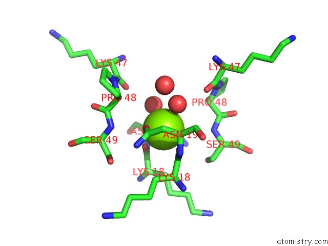
Mono view
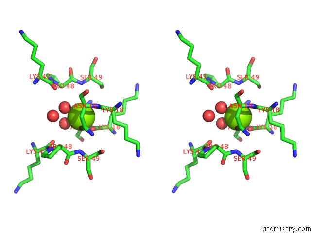
Stereo pair view
Mono view
Stereo pair view
A full contact list of Magnesium with other atoms in the Mg binding
site number 2 of X-Ray Structure of Homo Sapiens Protein FLJ36880 within 5.0Å range:
|
Magnesium binding site 3 out of 3 in 1saw
Go back to
Magnesium binding site 3 out
of 3 in the X-Ray Structure of Homo Sapiens Protein FLJ36880
Mono view
Stereo pair view
Mono view
Stereo pair view
A full contact list of Magnesium with other atoms in the Mg binding
site number 3 of X-Ray Structure of Homo Sapiens Protein FLJ36880 within 5.0Å range:
|
Reference:
B.A.Manjasetty,
F.H.Niesen,
H.Delbruck,
F.Gotz,
V.Sievert,
K.Bussow,
J.Behlke,
U.Heinemann.
X-Ray Structure of Fumarylacetoacetate Hydrolase Family Member Homo Sapiens FLJ36880. Biol.Chem. V. 385 935 2004.
ISSN: ISSN 1431-6730
PubMed: 15551868
DOI: 10.1515/BC.2004.122
Page generated: Tue Aug 13 13:42:17 2024
ISSN: ISSN 1431-6730
PubMed: 15551868
DOI: 10.1515/BC.2004.122
Last articles
Zn in 9J0NZn in 9J0O
Zn in 9J0P
Zn in 9FJX
Zn in 9EKB
Zn in 9C0F
Zn in 9CAH
Zn in 9CH0
Zn in 9CH3
Zn in 9CH1