Magnesium »
PDB 1s9d-1so5 »
1sd0 »
Magnesium in PDB 1sd0: Structure of Arginine Kinase C271A Mutant
Enzymatic activity of Structure of Arginine Kinase C271A Mutant
All present enzymatic activity of Structure of Arginine Kinase C271A Mutant:
2.7.3.3;
2.7.3.3;
Protein crystallography data
The structure of Structure of Arginine Kinase C271A Mutant, PDB code: 1sd0
was solved by
J.L.Gattis,
E.Ruben,
M.O.Fenley,
W.R.Ellington,
M.S.Chapman,
with X-Ray Crystallography technique. A brief refinement statistics is given in the table below:
| Resolution Low / High (Å) | 10.00 / 2.30 |
| Space group | P 21 21 21 |
| Cell size a, b, c (Å), α, β, γ (°) | 64.905, 71.255, 79.980, 90.00, 90.00, 90.00 |
| R / Rfree (%) | 20.9 / 23.6 |
Other elements in 1sd0:
The structure of Structure of Arginine Kinase C271A Mutant also contains other interesting chemical elements:
| Chlorine | (Cl) | 1 atom |
Magnesium Binding Sites:
The binding sites of Magnesium atom in the Structure of Arginine Kinase C271A Mutant
(pdb code 1sd0). This binding sites where shown within
5.0 Angstroms radius around Magnesium atom.
In total only one binding site of Magnesium was determined in the Structure of Arginine Kinase C271A Mutant, PDB code: 1sd0:
In total only one binding site of Magnesium was determined in the Structure of Arginine Kinase C271A Mutant, PDB code: 1sd0:
Magnesium binding site 1 out of 1 in 1sd0
Go back to
Magnesium binding site 1 out
of 1 in the Structure of Arginine Kinase C271A Mutant
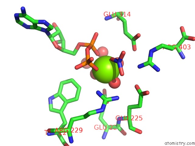
Mono view
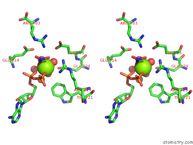
Stereo pair view
Mono view
Stereo pair view
A full contact list of Magnesium with other atoms in the Mg binding
site number 1 of Structure of Arginine Kinase C271A Mutant within 5.0Å range:
|
Reference:
J.L.Gattis,
E.Ruben,
M.O.Fenley,
W.R.Ellington,
M.S.Chapman.
The Active Site Cysteine of Arginine Kinase: Structural and Functional Analysis of Partially Active Mutants Biochemistry V. 43 8680 2004.
ISSN: ISSN 0006-2960
PubMed: 15236576
DOI: 10.1021/BI049793I
Page generated: Tue Aug 13 13:42:49 2024
ISSN: ISSN 0006-2960
PubMed: 15236576
DOI: 10.1021/BI049793I
Last articles
Zn in 9MJ5Zn in 9HNW
Zn in 9G0L
Zn in 9FNE
Zn in 9DZN
Zn in 9E0I
Zn in 9D32
Zn in 9DAK
Zn in 8ZXC
Zn in 8ZUF