Magnesium »
PDB 1s9d-1so5 »
1sjb »
Magnesium in PDB 1sjb: X-Ray Structure of O-Succinylbenzoate Synthase Complexed with O-Succinylbenzoic Acid
Protein crystallography data
The structure of X-Ray Structure of O-Succinylbenzoate Synthase Complexed with O-Succinylbenzoic Acid, PDB code: 1sjb
was solved by
J.B.Thoden,
E.A.Taylor-Ringia,
J.B.Garrett,
J.A.Gerlt,
H.M.Holden,
I.Rayment,
with X-Ray Crystallography technique. A brief refinement statistics is given in the table below:
| Resolution Low / High (Å) | 30.00 / 2.20 |
| Space group | H 3 2 |
| Cell size a, b, c (Å), α, β, γ (°) | 216.000, 216.000, 261.000, 90.00, 90.00, 120.00 |
| R / Rfree (%) | n/a / n/a |
Magnesium Binding Sites:
The binding sites of Magnesium atom in the X-Ray Structure of O-Succinylbenzoate Synthase Complexed with O-Succinylbenzoic Acid
(pdb code 1sjb). This binding sites where shown within
5.0 Angstroms radius around Magnesium atom.
In total 4 binding sites of Magnesium where determined in the X-Ray Structure of O-Succinylbenzoate Synthase Complexed with O-Succinylbenzoic Acid, PDB code: 1sjb:
Jump to Magnesium binding site number: 1; 2; 3; 4;
In total 4 binding sites of Magnesium where determined in the X-Ray Structure of O-Succinylbenzoate Synthase Complexed with O-Succinylbenzoic Acid, PDB code: 1sjb:
Jump to Magnesium binding site number: 1; 2; 3; 4;
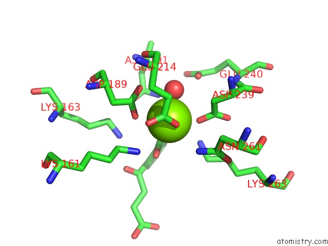
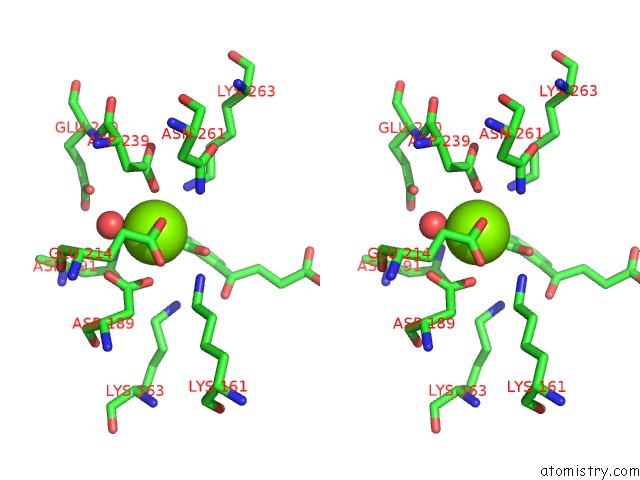
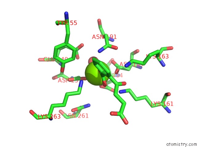
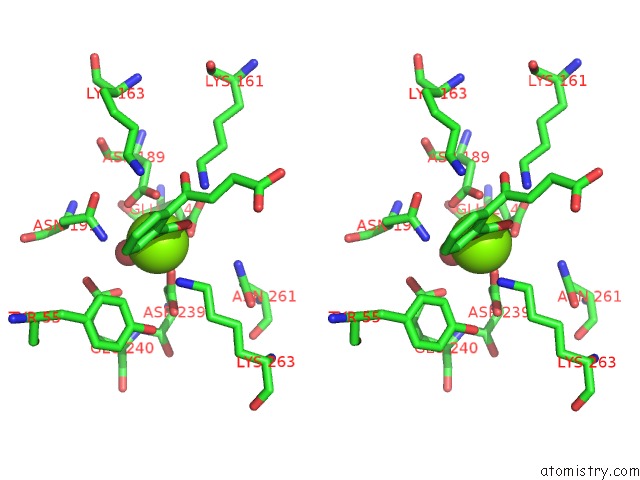
Magnesium binding site 1 out of 4 in 1sjb
Go back to
Magnesium binding site 1 out
of 4 in the X-Ray Structure of O-Succinylbenzoate Synthase Complexed with O-Succinylbenzoic Acid
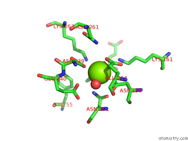
Mono view
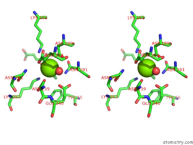
Stereo pair view
Mono view
Stereo pair view
A full contact list of Magnesium with other atoms in the Mg binding
site number 1 of X-Ray Structure of O-Succinylbenzoate Synthase Complexed with O-Succinylbenzoic Acid within 5.0Å range:
|
Magnesium binding site 2 out of 4 in 1sjb
Go back to
Magnesium binding site 2 out
of 4 in the X-Ray Structure of O-Succinylbenzoate Synthase Complexed with O-Succinylbenzoic Acid
Mono view
Stereo pair view
Mono view
Stereo pair view
A full contact list of Magnesium with other atoms in the Mg binding
site number 2 of X-Ray Structure of O-Succinylbenzoate Synthase Complexed with O-Succinylbenzoic Acid within 5.0Å range:
|
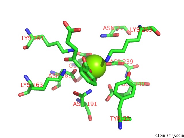
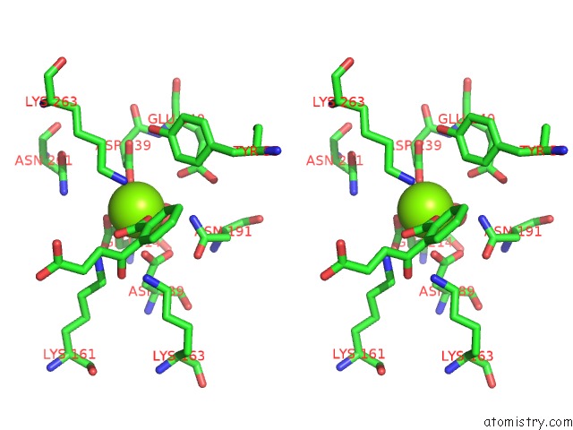
Magnesium binding site 3 out of 4 in 1sjb
Go back to
Magnesium binding site 3 out
of 4 in the X-Ray Structure of O-Succinylbenzoate Synthase Complexed with O-Succinylbenzoic Acid
Mono view
Stereo pair view
Mono view
Stereo pair view
A full contact list of Magnesium with other atoms in the Mg binding
site number 3 of X-Ray Structure of O-Succinylbenzoate Synthase Complexed with O-Succinylbenzoic Acid within 5.0Å range:
|
Magnesium binding site 4 out of 4 in 1sjb
Go back to
Magnesium binding site 4 out
of 4 in the X-Ray Structure of O-Succinylbenzoate Synthase Complexed with O-Succinylbenzoic Acid
Mono view
Stereo pair view
Mono view
Stereo pair view
A full contact list of Magnesium with other atoms in the Mg binding
site number 4 of X-Ray Structure of O-Succinylbenzoate Synthase Complexed with O-Succinylbenzoic Acid within 5.0Å range:
|
Reference:
J.B.Thoden,
E.A.Taylor-Ringia,
J.B.Garrett,
J.A.Gerlt,
H.M.Holden,
I.Rayment.
Evolution of Enzymatic Activity in the Enolase Superfamily: Structural Studies of the Promiscuous O-Succinylbenzoate Synthase From Amycolatopsis Biochemistry V. 43 5716 2004.
ISSN: ISSN 0006-2960
PubMed: 15134446
DOI: 10.1021/BI0497897
Page generated: Sun Aug 10 04:23:13 2025
ISSN: ISSN 0006-2960
PubMed: 15134446
DOI: 10.1021/BI0497897
Last articles
Mg in 4DSCMg in 4DS6
Mg in 4DRX
Mg in 4DR4
Mg in 4DR2
Mg in 4DR3
Mg in 4DR1
Mg in 4DPG
Mg in 4DQP
Mg in 4DQQ