Magnesium »
PDB 1s9d-1so5 »
1so4 »
Magnesium in PDB 1so4: Crystal Structure of K64A Mutant of 3-Keto-L-Gulonate 6- Phosphate Decarboxylase with Bound L-Threonohydroxamate 4- Phosphate
Protein crystallography data
The structure of Crystal Structure of K64A Mutant of 3-Keto-L-Gulonate 6- Phosphate Decarboxylase with Bound L-Threonohydroxamate 4- Phosphate, PDB code: 1so4
was solved by
E.L.Wise,
W.S.Yew,
J.A.Gerlt,
I.Rayment,
with X-Ray Crystallography technique. A brief refinement statistics is given in the table below:
| Resolution Low / High (Å) | 91.29 / 1.70 |
| Space group | C 1 2 1 |
| Cell size a, b, c (Å), α, β, γ (°) | 122.976, 41.900, 91.322, 90.00, 96.92, 90.00 |
| R / Rfree (%) | 16.2 / 20.5 |
Magnesium Binding Sites:
The binding sites of Magnesium atom in the Crystal Structure of K64A Mutant of 3-Keto-L-Gulonate 6- Phosphate Decarboxylase with Bound L-Threonohydroxamate 4- Phosphate
(pdb code 1so4). This binding sites where shown within
5.0 Angstroms radius around Magnesium atom.
In total 2 binding sites of Magnesium where determined in the Crystal Structure of K64A Mutant of 3-Keto-L-Gulonate 6- Phosphate Decarboxylase with Bound L-Threonohydroxamate 4- Phosphate, PDB code: 1so4:
Jump to Magnesium binding site number: 1; 2;
In total 2 binding sites of Magnesium where determined in the Crystal Structure of K64A Mutant of 3-Keto-L-Gulonate 6- Phosphate Decarboxylase with Bound L-Threonohydroxamate 4- Phosphate, PDB code: 1so4:
Jump to Magnesium binding site number: 1; 2;
Magnesium binding site 1 out of 2 in 1so4
Go back to
Magnesium binding site 1 out
of 2 in the Crystal Structure of K64A Mutant of 3-Keto-L-Gulonate 6- Phosphate Decarboxylase with Bound L-Threonohydroxamate 4- Phosphate
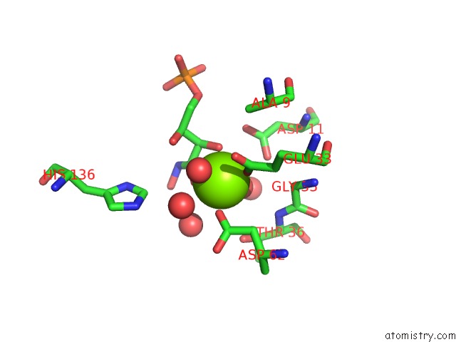
Mono view
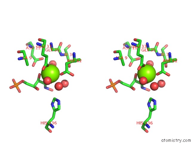
Stereo pair view
Mono view
Stereo pair view
A full contact list of Magnesium with other atoms in the Mg binding
site number 1 of Crystal Structure of K64A Mutant of 3-Keto-L-Gulonate 6- Phosphate Decarboxylase with Bound L-Threonohydroxamate 4- Phosphate within 5.0Å range:
|
Magnesium binding site 2 out of 2 in 1so4
Go back to
Magnesium binding site 2 out
of 2 in the Crystal Structure of K64A Mutant of 3-Keto-L-Gulonate 6- Phosphate Decarboxylase with Bound L-Threonohydroxamate 4- Phosphate
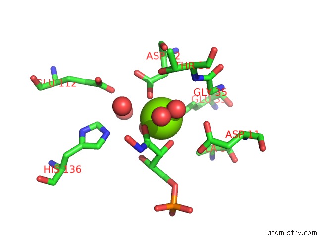
Mono view
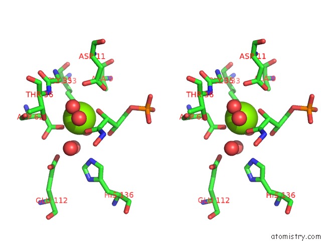
Stereo pair view
Mono view
Stereo pair view
A full contact list of Magnesium with other atoms in the Mg binding
site number 2 of Crystal Structure of K64A Mutant of 3-Keto-L-Gulonate 6- Phosphate Decarboxylase with Bound L-Threonohydroxamate 4- Phosphate within 5.0Å range:
|
Reference:
E.L.Wise,
W.S.Yew,
J.A.Gerlt,
I.Rayment.
Evolution of Enzymatic Activities in the Orotidine 5'-Monophosphate Decarboxylase Suprafamily: Crystallographic Evidence For A Proton Relay System in the Active Site of 3-Keto-L-Gulonate 6-Phosphate Decarboxylase(,) Biochemistry V. 43 6438 2004.
ISSN: ISSN 0006-2960
PubMed: 15157078
DOI: 10.1021/BI0497392
Page generated: Tue Aug 13 13:47:44 2024
ISSN: ISSN 0006-2960
PubMed: 15157078
DOI: 10.1021/BI0497392
Last articles
Zn in 9JYWZn in 9IR4
Zn in 9IR3
Zn in 9GMX
Zn in 9GMW
Zn in 9JEJ
Zn in 9ERF
Zn in 9ERE
Zn in 9EGV
Zn in 9EGW