Magnesium »
PDB 1so6-1t5s »
1sqk »
Magnesium in PDB 1sqk: Crystal Structure of Ciboulot in Complex with Skeletal Actin
Protein crystallography data
The structure of Crystal Structure of Ciboulot in Complex with Skeletal Actin, PDB code: 1sqk
was solved by
M.Hertzog,
C.Van Heijenoort,
D.Didry,
M.Gaudier,
B.Gigant,
J.Coutant,
G.Didelot,
T.Preat,
M.Knossow,
E.Guittet,
M.F.Carlier,
with X-Ray Crystallography technique. A brief refinement statistics is given in the table below:
| Resolution Low / High (Å) | 20.00 / 2.50 |
| Space group | P 21 21 21 |
| Cell size a, b, c (Å), α, β, γ (°) | 67.494, 75.198, 85.314, 90.00, 90.00, 90.00 |
| R / Rfree (%) | 21.9 / 27.8 |
Magnesium Binding Sites:
The binding sites of Magnesium atom in the Crystal Structure of Ciboulot in Complex with Skeletal Actin
(pdb code 1sqk). This binding sites where shown within
5.0 Angstroms radius around Magnesium atom.
In total only one binding site of Magnesium was determined in the Crystal Structure of Ciboulot in Complex with Skeletal Actin, PDB code: 1sqk:
In total only one binding site of Magnesium was determined in the Crystal Structure of Ciboulot in Complex with Skeletal Actin, PDB code: 1sqk:
Magnesium binding site 1 out of 1 in 1sqk
Go back to
Magnesium binding site 1 out
of 1 in the Crystal Structure of Ciboulot in Complex with Skeletal Actin
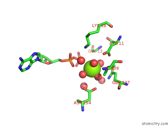
Mono view
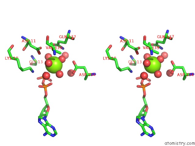
Stereo pair view
Mono view
Stereo pair view
A full contact list of Magnesium with other atoms in the Mg binding
site number 1 of Crystal Structure of Ciboulot in Complex with Skeletal Actin within 5.0Å range:
|
Reference:
M.Hertzog,
C.Van Heijenoort,
D.Didry,
M.Gaudier,
J.Coutant,
B.Gigant,
G.Didelot,
M.Knossow,
E.Guittet,
M.F.Carlier.
The Beta-Thymosin/WH2 Domain; Structural Basis For the Switch From Inhibition to Promotion of Actin Assembly Cell(Cambridge,Mass.) V. 117 611 2004.
ISSN: ISSN 0092-8674
PubMed: 15163409
DOI: 10.1016/S0092-8674(04)00403-9
Page generated: Tue Aug 13 14:18:12 2024
ISSN: ISSN 0092-8674
PubMed: 15163409
DOI: 10.1016/S0092-8674(04)00403-9
Last articles
Zn in 9J0NZn in 9J0O
Zn in 9J0P
Zn in 9FJX
Zn in 9EKB
Zn in 9C0F
Zn in 9CAH
Zn in 9CH0
Zn in 9CH3
Zn in 9CH1