Magnesium »
PDB 1so6-1t5s »
1sxj »
Magnesium in PDB 1sxj: Crystal Structure of the Eukaryotic Clamp Loader (Replication Factor C, Rfc) Bound to the Dna Sliding Clamp (Proliferating Cell Nuclear Antigen, Pcna)
Protein crystallography data
The structure of Crystal Structure of the Eukaryotic Clamp Loader (Replication Factor C, Rfc) Bound to the Dna Sliding Clamp (Proliferating Cell Nuclear Antigen, Pcna), PDB code: 1sxj
was solved by
G.D.Bowman,
M.O'donnell,
J.Kuriyan,
with X-Ray Crystallography technique. A brief refinement statistics is given in the table below:
| Resolution Low / High (Å) | 48.81 / 2.85 |
| Space group | P 21 21 21 |
| Cell size a, b, c (Å), α, β, γ (°) | 104.209, 110.481, 268.206, 90.00, 90.00, 90.00 |
| R / Rfree (%) | 25.1 / 30.6 |
Magnesium Binding Sites:
The binding sites of Magnesium atom in the Crystal Structure of the Eukaryotic Clamp Loader (Replication Factor C, Rfc) Bound to the Dna Sliding Clamp (Proliferating Cell Nuclear Antigen, Pcna)
(pdb code 1sxj). This binding sites where shown within
5.0 Angstroms radius around Magnesium atom.
In total 4 binding sites of Magnesium where determined in the Crystal Structure of the Eukaryotic Clamp Loader (Replication Factor C, Rfc) Bound to the Dna Sliding Clamp (Proliferating Cell Nuclear Antigen, Pcna), PDB code: 1sxj:
Jump to Magnesium binding site number: 1; 2; 3; 4;
In total 4 binding sites of Magnesium where determined in the Crystal Structure of the Eukaryotic Clamp Loader (Replication Factor C, Rfc) Bound to the Dna Sliding Clamp (Proliferating Cell Nuclear Antigen, Pcna), PDB code: 1sxj:
Jump to Magnesium binding site number: 1; 2; 3; 4;
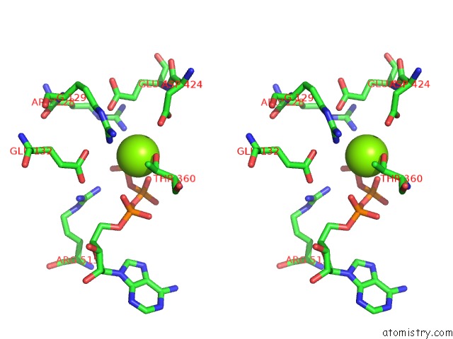
Magnesium binding site 1 out of 4 in 1sxj
Go back to
Magnesium binding site 1 out
of 4 in the Crystal Structure of the Eukaryotic Clamp Loader (Replication Factor C, Rfc) Bound to the Dna Sliding Clamp (Proliferating Cell Nuclear Antigen, Pcna)

Mono view
Stereo pair view
Mono view
Stereo pair view
A full contact list of Magnesium with other atoms in the Mg binding
site number 1 of Crystal Structure of the Eukaryotic Clamp Loader (Replication Factor C, Rfc) Bound to the Dna Sliding Clamp (Proliferating Cell Nuclear Antigen, Pcna) within 5.0Å range:
|
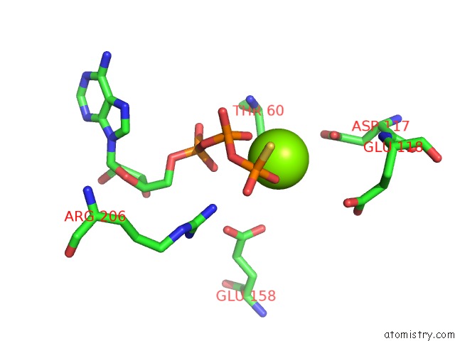
Magnesium binding site 2 out of 4 in 1sxj
Go back to
Magnesium binding site 2 out
of 4 in the Crystal Structure of the Eukaryotic Clamp Loader (Replication Factor C, Rfc) Bound to the Dna Sliding Clamp (Proliferating Cell Nuclear Antigen, Pcna)

Mono view

Stereo pair view
Mono view
Stereo pair view
A full contact list of Magnesium with other atoms in the Mg binding
site number 2 of Crystal Structure of the Eukaryotic Clamp Loader (Replication Factor C, Rfc) Bound to the Dna Sliding Clamp (Proliferating Cell Nuclear Antigen, Pcna) within 5.0Å range:
|
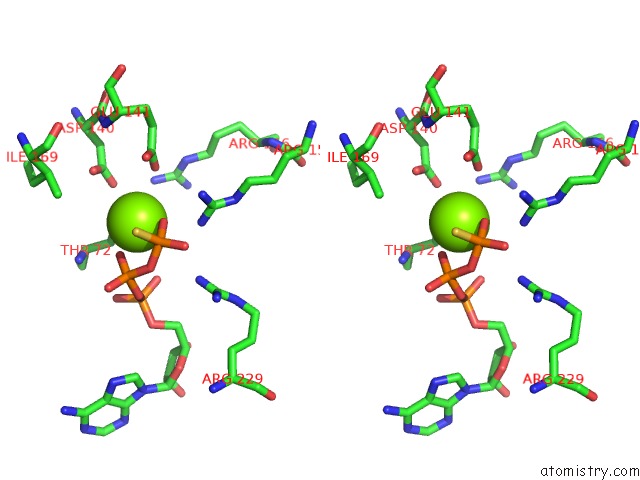
Magnesium binding site 3 out of 4 in 1sxj
Go back to
Magnesium binding site 3 out
of 4 in the Crystal Structure of the Eukaryotic Clamp Loader (Replication Factor C, Rfc) Bound to the Dna Sliding Clamp (Proliferating Cell Nuclear Antigen, Pcna)
Mono view

Stereo pair view
Mono view
Stereo pair view
A full contact list of Magnesium with other atoms in the Mg binding
site number 3 of Crystal Structure of the Eukaryotic Clamp Loader (Replication Factor C, Rfc) Bound to the Dna Sliding Clamp (Proliferating Cell Nuclear Antigen, Pcna) within 5.0Å range:
|
Magnesium binding site 4 out of 4 in 1sxj
Go back to
Magnesium binding site 4 out
of 4 in the Crystal Structure of the Eukaryotic Clamp Loader (Replication Factor C, Rfc) Bound to the Dna Sliding Clamp (Proliferating Cell Nuclear Antigen, Pcna)

Mono view
Stereo pair view
Mono view
Stereo pair view
A full contact list of Magnesium with other atoms in the Mg binding
site number 4 of Crystal Structure of the Eukaryotic Clamp Loader (Replication Factor C, Rfc) Bound to the Dna Sliding Clamp (Proliferating Cell Nuclear Antigen, Pcna) within 5.0Å range:
|
Reference:
G.D.Bowman,
M.O'donnell,
J.Kuriyan.
Structural Analysis of A Eukaryotic Sliding Dna Clamp-Clamp Loader Complex. Nature V. 429 724 2004.
ISSN: ISSN 0028-0836
PubMed: 15201901
DOI: 10.1038/NATURE02585
Page generated: Tue Aug 13 14:22:41 2024
ISSN: ISSN 0028-0836
PubMed: 15201901
DOI: 10.1038/NATURE02585
Last articles
Zn in 9MJ5Zn in 9HNW
Zn in 9G0L
Zn in 9FNE
Zn in 9DZN
Zn in 9E0I
Zn in 9D32
Zn in 9DAK
Zn in 8ZXC
Zn in 8ZUF