Magnesium »
PDB 1t5t-1tkd »
1tj5 »
Magnesium in PDB 1tj5: X-Ray Structure of the Sucrose-Phosphatase (Spp) From Synechocystis Sp. PCC6803 in Complex with Sucrose and Phosphate
Enzymatic activity of X-Ray Structure of the Sucrose-Phosphatase (Spp) From Synechocystis Sp. PCC6803 in Complex with Sucrose and Phosphate
All present enzymatic activity of X-Ray Structure of the Sucrose-Phosphatase (Spp) From Synechocystis Sp. PCC6803 in Complex with Sucrose and Phosphate:
3.1.3.24;
3.1.3.24;
Protein crystallography data
The structure of X-Ray Structure of the Sucrose-Phosphatase (Spp) From Synechocystis Sp. PCC6803 in Complex with Sucrose and Phosphate, PDB code: 1tj5
was solved by
S.Fieulaine,
J.E.Lunn,
F.Borel,
J.-L.Ferrer,
with X-Ray Crystallography technique. A brief refinement statistics is given in the table below:
| Resolution Low / High (Å) | 50.00 / 2.20 |
| Space group | P 65 2 2 |
| Cell size a, b, c (Å), α, β, γ (°) | 68.750, 68.750, 268.380, 90.00, 90.00, 120.00 |
| R / Rfree (%) | 18.9 / 22 |
Magnesium Binding Sites:
The binding sites of Magnesium atom in the X-Ray Structure of the Sucrose-Phosphatase (Spp) From Synechocystis Sp. PCC6803 in Complex with Sucrose and Phosphate
(pdb code 1tj5). This binding sites where shown within
5.0 Angstroms radius around Magnesium atom.
In total 2 binding sites of Magnesium where determined in the X-Ray Structure of the Sucrose-Phosphatase (Spp) From Synechocystis Sp. PCC6803 in Complex with Sucrose and Phosphate, PDB code: 1tj5:
Jump to Magnesium binding site number: 1; 2;
In total 2 binding sites of Magnesium where determined in the X-Ray Structure of the Sucrose-Phosphatase (Spp) From Synechocystis Sp. PCC6803 in Complex with Sucrose and Phosphate, PDB code: 1tj5:
Jump to Magnesium binding site number: 1; 2;
Magnesium binding site 1 out of 2 in 1tj5
Go back to
Magnesium binding site 1 out
of 2 in the X-Ray Structure of the Sucrose-Phosphatase (Spp) From Synechocystis Sp. PCC6803 in Complex with Sucrose and Phosphate
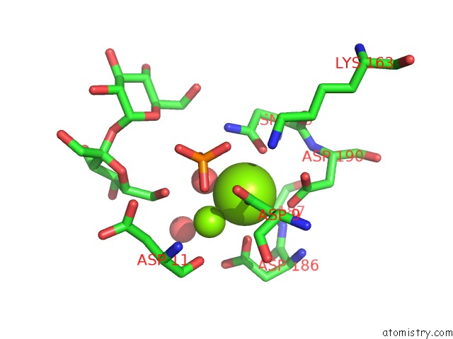
Mono view
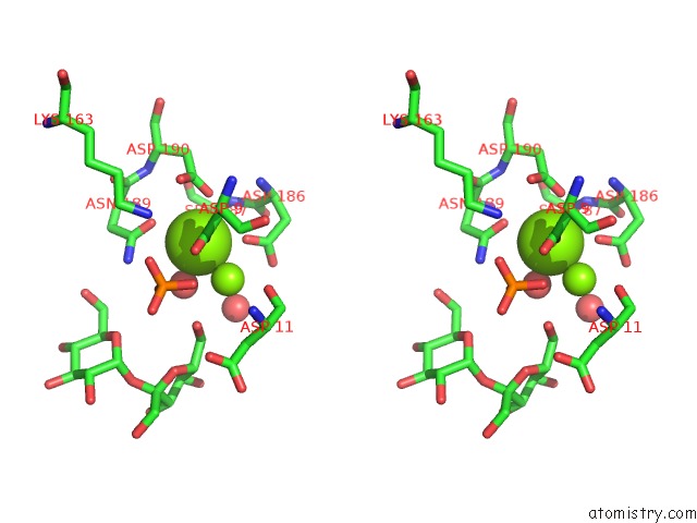
Stereo pair view
Mono view
Stereo pair view
A full contact list of Magnesium with other atoms in the Mg binding
site number 1 of X-Ray Structure of the Sucrose-Phosphatase (Spp) From Synechocystis Sp. PCC6803 in Complex with Sucrose and Phosphate within 5.0Å range:
|
Magnesium binding site 2 out of 2 in 1tj5
Go back to
Magnesium binding site 2 out
of 2 in the X-Ray Structure of the Sucrose-Phosphatase (Spp) From Synechocystis Sp. PCC6803 in Complex with Sucrose and Phosphate
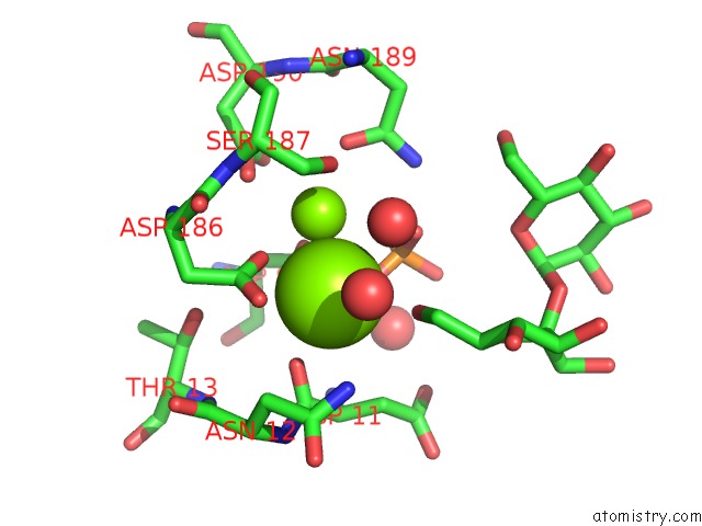
Mono view
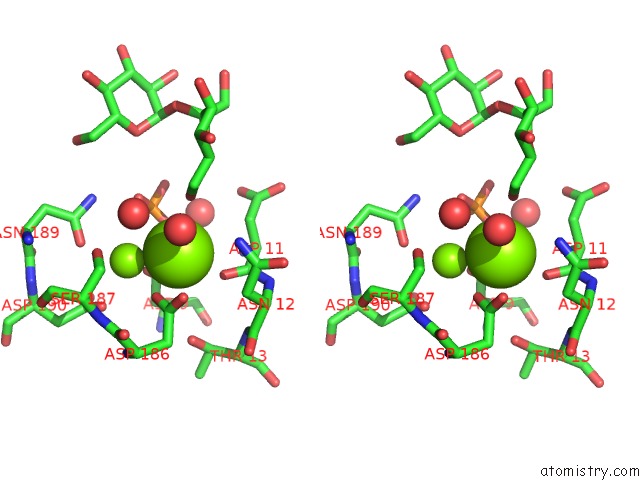
Stereo pair view
Mono view
Stereo pair view
A full contact list of Magnesium with other atoms in the Mg binding
site number 2 of X-Ray Structure of the Sucrose-Phosphatase (Spp) From Synechocystis Sp. PCC6803 in Complex with Sucrose and Phosphate within 5.0Å range:
|
Reference:
S.Fieulaine,
J.E.Lunn,
F.Borel,
J.-L.Ferrer.
The Structure of A Cyanobacterial Sucrose-Phosphatase Reveals the Sugar Tongs That Release Free Sucrose in the Cell. Plant Cell V. 17 2049 2005.
ISSN: ISSN 1040-4651
PubMed: 15937230
DOI: 10.1105/TPC.105.031229
Page generated: Tue Aug 13 14:33:28 2024
ISSN: ISSN 1040-4651
PubMed: 15937230
DOI: 10.1105/TPC.105.031229
Last articles
Zn in 9J0NZn in 9J0O
Zn in 9J0P
Zn in 9FJX
Zn in 9EKB
Zn in 9C0F
Zn in 9CAH
Zn in 9CH0
Zn in 9CH3
Zn in 9CH1