Magnesium »
PDB 1tkk-1u3f »
1tzf »
Magnesium in PDB 1tzf: X-Ray Crystal Structure of Alpha-D-Glucose-1-Phosphate Cytidylyltransferase From Salmonella Typhi
Enzymatic activity of X-Ray Crystal Structure of Alpha-D-Glucose-1-Phosphate Cytidylyltransferase From Salmonella Typhi
All present enzymatic activity of X-Ray Crystal Structure of Alpha-D-Glucose-1-Phosphate Cytidylyltransferase From Salmonella Typhi:
2.7.7.33;
2.7.7.33;
Protein crystallography data
The structure of X-Ray Crystal Structure of Alpha-D-Glucose-1-Phosphate Cytidylyltransferase From Salmonella Typhi, PDB code: 1tzf
was solved by
N.M.Koropatkin,
H.M.Holden,
with X-Ray Crystallography technique. A brief refinement statistics is given in the table below:
| Resolution Low / High (Å) | 50.00 / 2.10 |
| Space group | P 63 2 2 |
| Cell size a, b, c (Å), α, β, γ (°) | 88.500, 88.500, 162.300, 90.00, 90.00, 120.00 |
| R / Rfree (%) | n/a / n/a |
Magnesium Binding Sites:
The binding sites of Magnesium atom in the X-Ray Crystal Structure of Alpha-D-Glucose-1-Phosphate Cytidylyltransferase From Salmonella Typhi
(pdb code 1tzf). This binding sites where shown within
5.0 Angstroms radius around Magnesium atom.
In total only one binding site of Magnesium was determined in the X-Ray Crystal Structure of Alpha-D-Glucose-1-Phosphate Cytidylyltransferase From Salmonella Typhi, PDB code: 1tzf:
In total only one binding site of Magnesium was determined in the X-Ray Crystal Structure of Alpha-D-Glucose-1-Phosphate Cytidylyltransferase From Salmonella Typhi, PDB code: 1tzf:
Magnesium binding site 1 out of 1 in 1tzf
Go back to
Magnesium binding site 1 out
of 1 in the X-Ray Crystal Structure of Alpha-D-Glucose-1-Phosphate Cytidylyltransferase From Salmonella Typhi
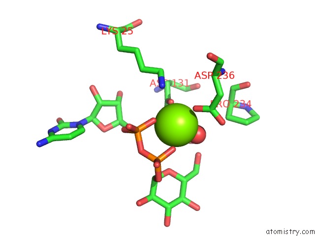
Mono view
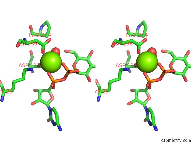
Stereo pair view
Mono view
Stereo pair view
A full contact list of Magnesium with other atoms in the Mg binding
site number 1 of X-Ray Crystal Structure of Alpha-D-Glucose-1-Phosphate Cytidylyltransferase From Salmonella Typhi within 5.0Å range:
|
Reference:
N.M.Koropatkin,
H.M.Holden.
Molecular Structure of Alpha-D-Glucose-1-Phosphate Cytidylyltransferase From Salmonella Typhi J.Biol.Chem. V. 279 44023 2004.
ISSN: ISSN 0021-9258
PubMed: 15292268
DOI: 10.1074/JBC.M407755200
Page generated: Tue Aug 13 14:40:48 2024
ISSN: ISSN 0021-9258
PubMed: 15292268
DOI: 10.1074/JBC.M407755200
Last articles
Zn in 9J0NZn in 9J0O
Zn in 9J0P
Zn in 9FJX
Zn in 9EKB
Zn in 9C0F
Zn in 9CAH
Zn in 9CH0
Zn in 9CH3
Zn in 9CH1