Magnesium »
PDB 1tkk-1u3f »
1u0h »
Magnesium in PDB 1u0h: Structural Basis For the Inhibition of Mammalian Adenylyl Cyclase By Mant-Gtp
Enzymatic activity of Structural Basis For the Inhibition of Mammalian Adenylyl Cyclase By Mant-Gtp
All present enzymatic activity of Structural Basis For the Inhibition of Mammalian Adenylyl Cyclase By Mant-Gtp:
4.6.1.1;
4.6.1.1;
Protein crystallography data
The structure of Structural Basis For the Inhibition of Mammalian Adenylyl Cyclase By Mant-Gtp, PDB code: 1u0h
was solved by
T.C.Mou,
A.Gille,
R.J.Seifert,
S.R.Sprang,
with X-Ray Crystallography technique. A brief refinement statistics is given in the table below:
| Resolution Low / High (Å) | 15.00 / 2.90 |
| Space group | P 21 21 2 |
| Cell size a, b, c (Å), α, β, γ (°) | 118.400, 133.000, 70.400, 90.00, 90.00, 90.00 |
| R / Rfree (%) | 24.5 / 28 |
Other elements in 1u0h:
The structure of Structural Basis For the Inhibition of Mammalian Adenylyl Cyclase By Mant-Gtp also contains other interesting chemical elements:
| Chlorine | (Cl) | 1 atom |
Magnesium Binding Sites:
The binding sites of Magnesium atom in the Structural Basis For the Inhibition of Mammalian Adenylyl Cyclase By Mant-Gtp
(pdb code 1u0h). This binding sites where shown within
5.0 Angstroms radius around Magnesium atom.
In total 3 binding sites of Magnesium where determined in the Structural Basis For the Inhibition of Mammalian Adenylyl Cyclase By Mant-Gtp, PDB code: 1u0h:
Jump to Magnesium binding site number: 1; 2; 3;
In total 3 binding sites of Magnesium where determined in the Structural Basis For the Inhibition of Mammalian Adenylyl Cyclase By Mant-Gtp, PDB code: 1u0h:
Jump to Magnesium binding site number: 1; 2; 3;
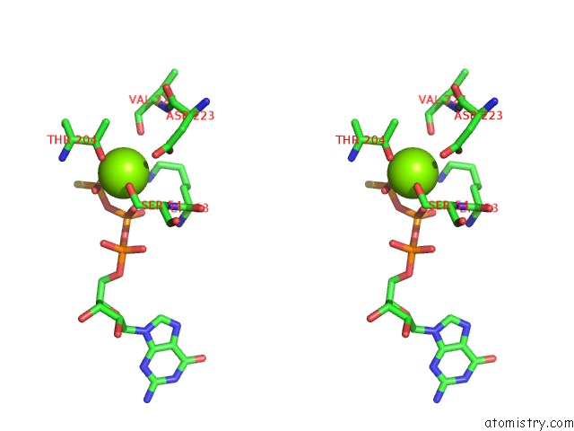
Magnesium binding site 1 out of 3 in 1u0h
Go back to
Magnesium binding site 1 out
of 3 in the Structural Basis For the Inhibition of Mammalian Adenylyl Cyclase By Mant-Gtp
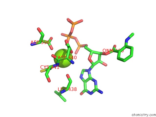
Mono view
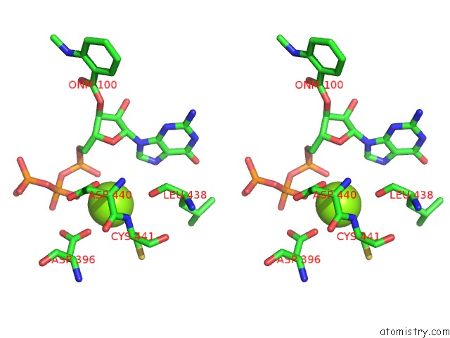
Stereo pair view
Mono view
Stereo pair view
A full contact list of Magnesium with other atoms in the Mg binding
site number 1 of Structural Basis For the Inhibition of Mammalian Adenylyl Cyclase By Mant-Gtp within 5.0Å range:
|
Magnesium binding site 2 out of 3 in 1u0h
Go back to
Magnesium binding site 2 out
of 3 in the Structural Basis For the Inhibition of Mammalian Adenylyl Cyclase By Mant-Gtp
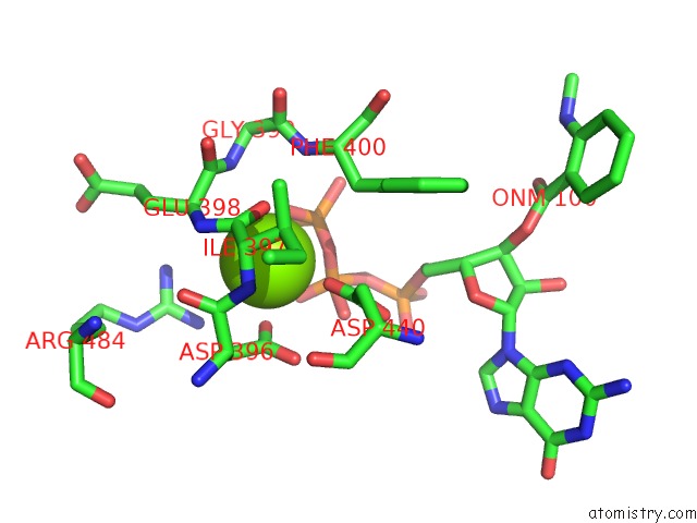
Mono view
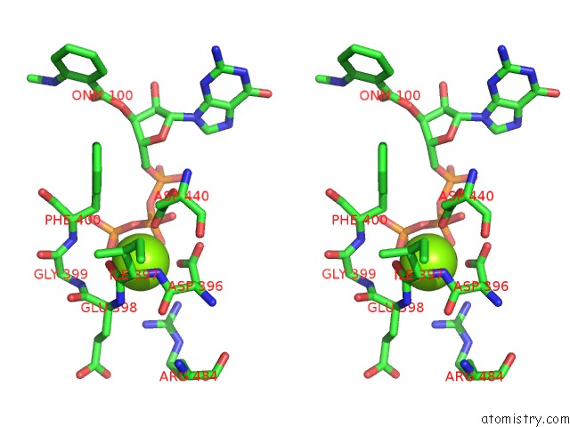
Stereo pair view
Mono view
Stereo pair view
A full contact list of Magnesium with other atoms in the Mg binding
site number 2 of Structural Basis For the Inhibition of Mammalian Adenylyl Cyclase By Mant-Gtp within 5.0Å range:
|
Magnesium binding site 3 out of 3 in 1u0h
Go back to
Magnesium binding site 3 out
of 3 in the Structural Basis For the Inhibition of Mammalian Adenylyl Cyclase By Mant-Gtp

Mono view
Stereo pair view
Mono view
Stereo pair view
A full contact list of Magnesium with other atoms in the Mg binding
site number 3 of Structural Basis For the Inhibition of Mammalian Adenylyl Cyclase By Mant-Gtp within 5.0Å range:
|
Reference:
T.C.Mou,
A.Gille,
D.A.Fancy,
R.Seifert,
S.R.Sprang.
Structural Basis For the Inhibition of Mammalian Membrane Adenylyl Cyclase By 2 '(3')-O-(N-Methylanthraniloyl)-Guanosine 5 '-Triphosphate. J.Biol.Chem. V. 280 7253 2005.
ISSN: ISSN 0021-9258
PubMed: 15591060
DOI: 10.1074/JBC.M409076200
Page generated: Tue Aug 13 14:41:19 2024
ISSN: ISSN 0021-9258
PubMed: 15591060
DOI: 10.1074/JBC.M409076200
Last articles
F in 7NTHF in 7NTI
F in 7NPC
F in 7NRG
F in 7NR5
F in 7NQS
F in 7NOS
F in 7NP5
F in 7NDV
F in 7NP6