Magnesium »
PDB 1u3g-1uet »
1u48 »
Magnesium in PDB 1u48: Extension of A Cytosine-8-Oxoguanine Base Pair
Enzymatic activity of Extension of A Cytosine-8-Oxoguanine Base Pair
All present enzymatic activity of Extension of A Cytosine-8-Oxoguanine Base Pair:
2.7.7.7;
2.7.7.7;
Protein crystallography data
The structure of Extension of A Cytosine-8-Oxoguanine Base Pair, PDB code: 1u48
was solved by
G.W.Hsu,
M.Ober,
T.Carell,
L.S.Beese,
with X-Ray Crystallography technique. A brief refinement statistics is given in the table below:
| Resolution Low / High (Å) | 40.78 / 2.10 |
| Space group | P 21 21 21 |
| Cell size a, b, c (Å), α, β, γ (°) | 87.342, 93.265, 106.084, 90.00, 90.00, 90.00 |
| R / Rfree (%) | 20.5 / 23.9 |
Magnesium Binding Sites:
The binding sites of Magnesium atom in the Extension of A Cytosine-8-Oxoguanine Base Pair
(pdb code 1u48). This binding sites where shown within
5.0 Angstroms radius around Magnesium atom.
In total only one binding site of Magnesium was determined in the Extension of A Cytosine-8-Oxoguanine Base Pair, PDB code: 1u48:
In total only one binding site of Magnesium was determined in the Extension of A Cytosine-8-Oxoguanine Base Pair, PDB code: 1u48:
Magnesium binding site 1 out of 1 in 1u48
Go back to
Magnesium binding site 1 out
of 1 in the Extension of A Cytosine-8-Oxoguanine Base Pair
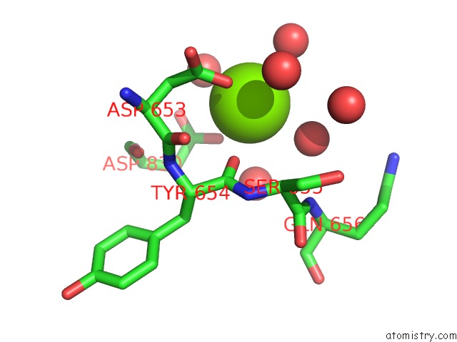
Mono view
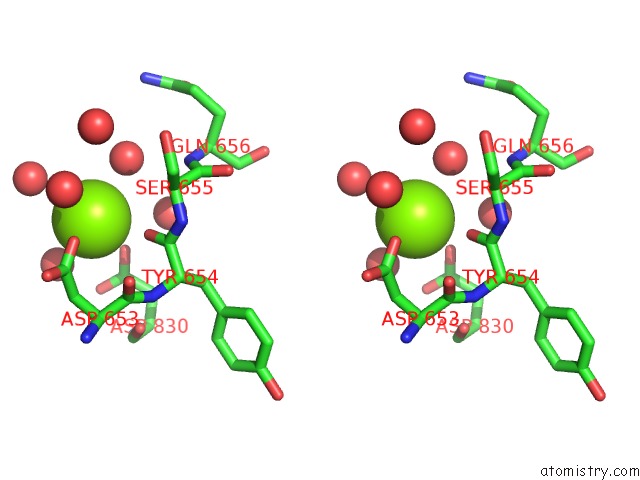
Stereo pair view
Mono view
Stereo pair view
A full contact list of Magnesium with other atoms in the Mg binding
site number 1 of Extension of A Cytosine-8-Oxoguanine Base Pair within 5.0Å range:
|
Reference:
G.W.Hsu,
M.Ober,
T.Carell,
L.S.Beese.
Error-Prone Replication of Oxidatively Damaged Dna By A High-Fidelity Dna Polymerase. Nature V. 431 217 2004.
ISSN: ISSN 0028-0836
PubMed: 15322558
DOI: 10.1038/NATURE02908
Page generated: Sun Aug 10 05:01:37 2025
ISSN: ISSN 0028-0836
PubMed: 15322558
DOI: 10.1038/NATURE02908
Last articles
Mg in 4EN4Mg in 4EM4
Mg in 4EMW
Mg in 4ELT
Mg in 4ELU
Mg in 4ELV
Mg in 4EM3
Mg in 4EKD
Mg in 4EHU
Mg in 4EHT