Magnesium »
PDB 1u3g-1uet »
1u9i »
Magnesium in PDB 1u9i: Crystal Structure of Circadian Clock Protein Kaic with Phosphorylation Sites
Protein crystallography data
The structure of Crystal Structure of Circadian Clock Protein Kaic with Phosphorylation Sites, PDB code: 1u9i
was solved by
Y.Xu,
T.Mori,
R.Pattanayek,
S.Pattanayek,
M.Egli,
C.H.Johnson,
with X-Ray Crystallography technique. A brief refinement statistics is given in the table below:
| Resolution Low / High (Å) | 30.00 / 2.80 |
| Space group | P 21 21 21 |
| Cell size a, b, c (Å), α, β, γ (°) | 132.873, 135.576, 204.951, 90.00, 90.00, 90.00 |
| R / Rfree (%) | 25 / 29 |
Magnesium Binding Sites:
The binding sites of Magnesium atom in the Crystal Structure of Circadian Clock Protein Kaic with Phosphorylation Sites
(pdb code 1u9i). This binding sites where shown within
5.0 Angstroms radius around Magnesium atom.
In total 6 binding sites of Magnesium where determined in the Crystal Structure of Circadian Clock Protein Kaic with Phosphorylation Sites, PDB code: 1u9i:
Jump to Magnesium binding site number: 1; 2; 3; 4; 5; 6;
In total 6 binding sites of Magnesium where determined in the Crystal Structure of Circadian Clock Protein Kaic with Phosphorylation Sites, PDB code: 1u9i:
Jump to Magnesium binding site number: 1; 2; 3; 4; 5; 6;
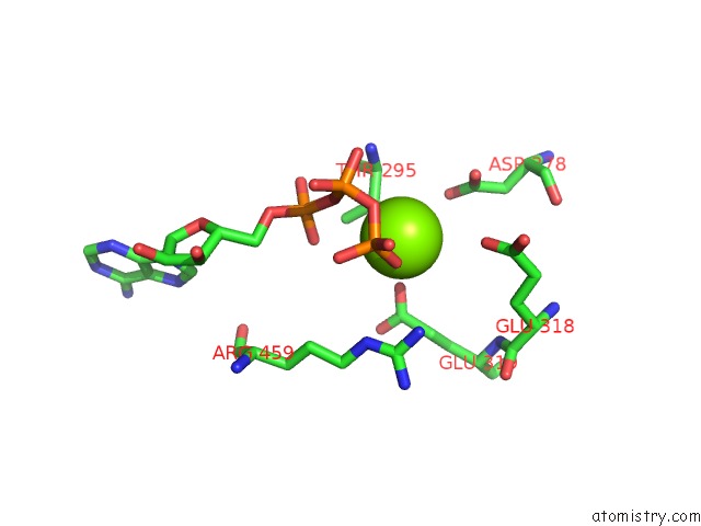
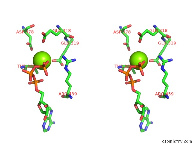
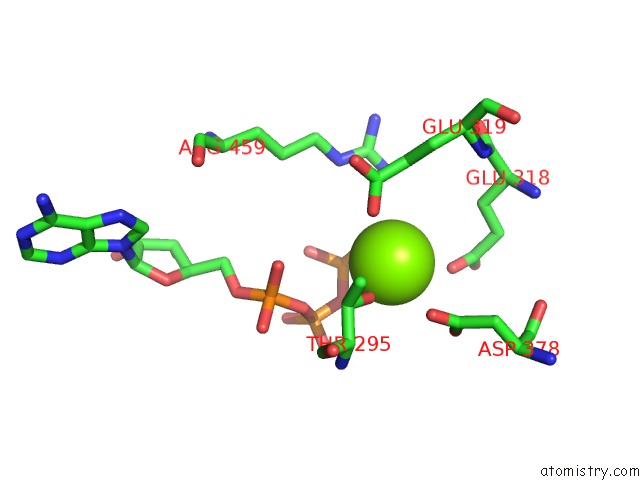
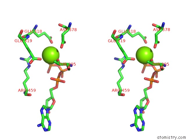
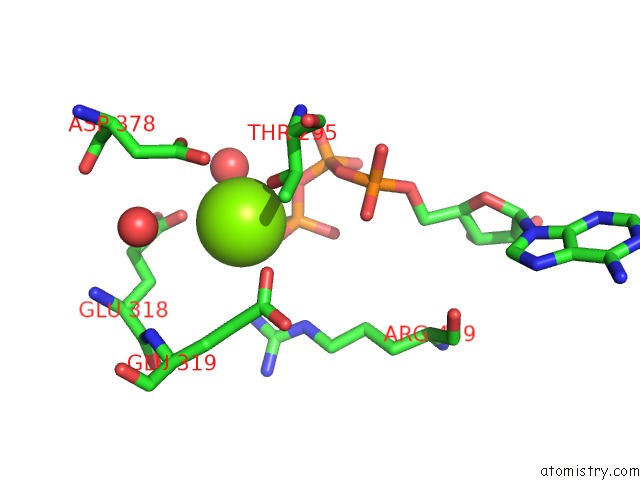
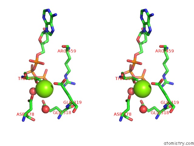
Magnesium binding site 1 out of 6 in 1u9i
Go back to
Magnesium binding site 1 out
of 6 in the Crystal Structure of Circadian Clock Protein Kaic with Phosphorylation Sites
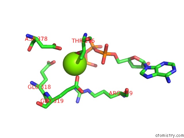
Mono view
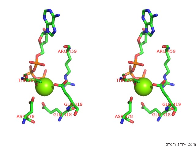
Stereo pair view
Mono view
Stereo pair view
A full contact list of Magnesium with other atoms in the Mg binding
site number 1 of Crystal Structure of Circadian Clock Protein Kaic with Phosphorylation Sites within 5.0Å range:
|
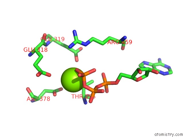
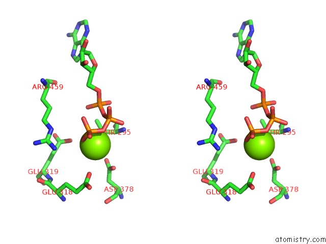
Magnesium binding site 2 out of 6 in 1u9i
Go back to
Magnesium binding site 2 out
of 6 in the Crystal Structure of Circadian Clock Protein Kaic with Phosphorylation Sites
Mono view
Stereo pair view
Mono view
Stereo pair view
A full contact list of Magnesium with other atoms in the Mg binding
site number 2 of Crystal Structure of Circadian Clock Protein Kaic with Phosphorylation Sites within 5.0Å range:
|
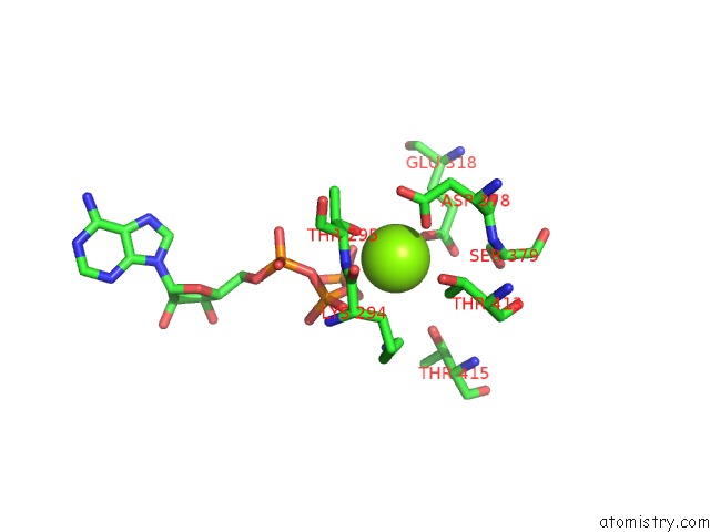
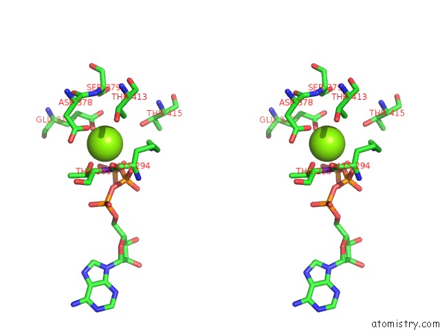
Magnesium binding site 3 out of 6 in 1u9i
Go back to
Magnesium binding site 3 out
of 6 in the Crystal Structure of Circadian Clock Protein Kaic with Phosphorylation Sites
Mono view
Stereo pair view
Mono view
Stereo pair view
A full contact list of Magnesium with other atoms in the Mg binding
site number 3 of Crystal Structure of Circadian Clock Protein Kaic with Phosphorylation Sites within 5.0Å range:
|
Magnesium binding site 4 out of 6 in 1u9i
Go back to
Magnesium binding site 4 out
of 6 in the Crystal Structure of Circadian Clock Protein Kaic with Phosphorylation Sites
Mono view
Stereo pair view
Mono view
Stereo pair view
A full contact list of Magnesium with other atoms in the Mg binding
site number 4 of Crystal Structure of Circadian Clock Protein Kaic with Phosphorylation Sites within 5.0Å range:
|
Magnesium binding site 5 out of 6 in 1u9i
Go back to
Magnesium binding site 5 out
of 6 in the Crystal Structure of Circadian Clock Protein Kaic with Phosphorylation Sites
Mono view
Stereo pair view
Mono view
Stereo pair view
A full contact list of Magnesium with other atoms in the Mg binding
site number 5 of Crystal Structure of Circadian Clock Protein Kaic with Phosphorylation Sites within 5.0Å range:
|
Magnesium binding site 6 out of 6 in 1u9i
Go back to
Magnesium binding site 6 out
of 6 in the Crystal Structure of Circadian Clock Protein Kaic with Phosphorylation Sites
Mono view
Stereo pair view
Mono view
Stereo pair view
A full contact list of Magnesium with other atoms in the Mg binding
site number 6 of Crystal Structure of Circadian Clock Protein Kaic with Phosphorylation Sites within 5.0Å range:
|
Reference:
Y.Xu,
T.Mori,
R.Pattanayek,
S.Pattanayek,
M.Egli,
C.H.Johnson.
Identification of Key Phosphorylation Sites in the Circadian Clock Protein Kaic By Crystallographic and Mutagenetic Analyses Proc.Natl.Acad.Sci.Usa V. 101 13933 2004.
ISSN: ISSN 0027-8424
PubMed: 15347809
DOI: 10.1073/PNAS.0404768101
Page generated: Tue Aug 13 14:47:39 2024
ISSN: ISSN 0027-8424
PubMed: 15347809
DOI: 10.1073/PNAS.0404768101
Last articles
F in 7L8JF in 7L8I
F in 7L7N
F in 7L8H
F in 7L7L
F in 7L7P
F in 7L7O
F in 7L5E
F in 7L72
F in 7L5P