Magnesium »
PDB 1u3g-1uet »
1uet »
Magnesium in PDB 1uet: Divergent Evolutions of Trinucleotide Polymerization Revealed By An Archaeal Cca-Adding Enzyme Structure
Enzymatic activity of Divergent Evolutions of Trinucleotide Polymerization Revealed By An Archaeal Cca-Adding Enzyme Structure
All present enzymatic activity of Divergent Evolutions of Trinucleotide Polymerization Revealed By An Archaeal Cca-Adding Enzyme Structure:
2.7.7.25;
2.7.7.25;
Protein crystallography data
The structure of Divergent Evolutions of Trinucleotide Polymerization Revealed By An Archaeal Cca-Adding Enzyme Structure, PDB code: 1uet
was solved by
O.Nureki,
Riken Structural Genomics/Proteomics Initiative (Rsgi),
with X-Ray Crystallography technique. A brief refinement statistics is given in the table below:
| Resolution Low / High (Å) | 48.02 / 2.00 |
| Space group | C 1 2 1 |
| Cell size a, b, c (Å), α, β, γ (°) | 87.351, 76.548, 76.771, 90.00, 98.33, 90.00 |
| R / Rfree (%) | 19.8 / 24.2 |
Other elements in 1uet:
The structure of Divergent Evolutions of Trinucleotide Polymerization Revealed By An Archaeal Cca-Adding Enzyme Structure also contains other interesting chemical elements:
| Calcium | (Ca) | 3 atoms |
Magnesium Binding Sites:
The binding sites of Magnesium atom in the Divergent Evolutions of Trinucleotide Polymerization Revealed By An Archaeal Cca-Adding Enzyme Structure
(pdb code 1uet). This binding sites where shown within
5.0 Angstroms radius around Magnesium atom.
In total only one binding site of Magnesium was determined in the Divergent Evolutions of Trinucleotide Polymerization Revealed By An Archaeal Cca-Adding Enzyme Structure, PDB code: 1uet:
In total only one binding site of Magnesium was determined in the Divergent Evolutions of Trinucleotide Polymerization Revealed By An Archaeal Cca-Adding Enzyme Structure, PDB code: 1uet:
Magnesium binding site 1 out of 1 in 1uet
Go back to
Magnesium binding site 1 out
of 1 in the Divergent Evolutions of Trinucleotide Polymerization Revealed By An Archaeal Cca-Adding Enzyme Structure
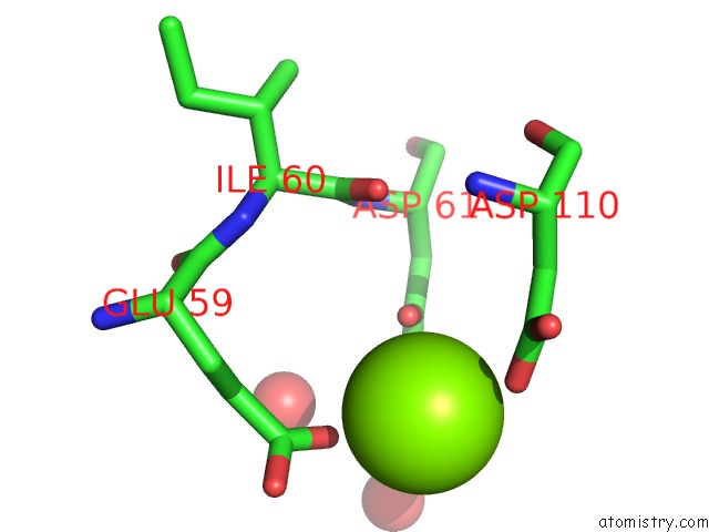
Mono view
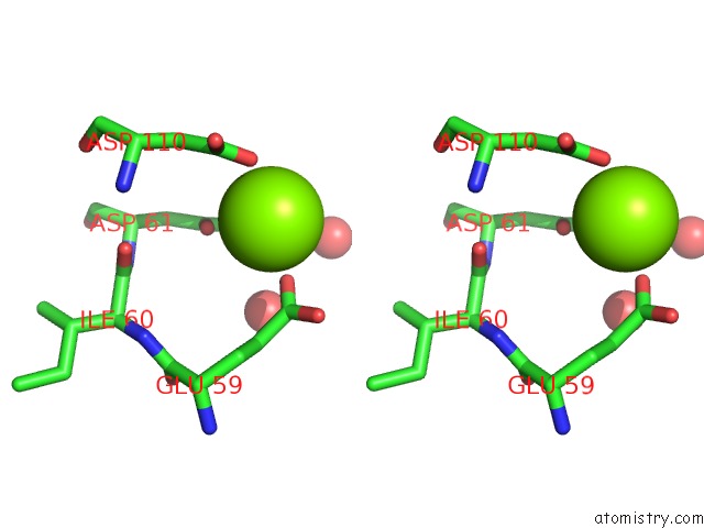
Stereo pair view
Mono view
Stereo pair view
A full contact list of Magnesium with other atoms in the Mg binding
site number 1 of Divergent Evolutions of Trinucleotide Polymerization Revealed By An Archaeal Cca-Adding Enzyme Structure within 5.0Å range:
|
Reference:
M.Okabe,
K.Tomita,
R.Ishitani,
R.Ishii,
N.Takeuchi,
F.Arisaka,
O.Nureki,
S.Yokoyama.
Divergent Evolutions of Trinucleotide Polymerization Revealed By An Archaeal Cca-Adding Enzyme Structure. Embo J. V. 22 5918 2003.
ISSN: ISSN 0261-4189
PubMed: 14592988
DOI: 10.1093/EMBOJ/CDG563
Page generated: Tue Aug 13 14:50:33 2024
ISSN: ISSN 0261-4189
PubMed: 14592988
DOI: 10.1093/EMBOJ/CDG563
Last articles
Zn in 9JYWZn in 9IR4
Zn in 9IR3
Zn in 9GMX
Zn in 9GMW
Zn in 9JEJ
Zn in 9ERF
Zn in 9ERE
Zn in 9EGV
Zn in 9EGW