Magnesium »
PDB 1v5g-1vq4 »
1v8j »
Magnesium in PDB 1v8j: The Crystal Structure of the Minimal Functional Domain of the Microtubule Destabilizer KIF2C Complexed with Mg-Adp
Protein crystallography data
The structure of The Crystal Structure of the Minimal Functional Domain of the Microtubule Destabilizer KIF2C Complexed with Mg-Adp, PDB code: 1v8j
was solved by
T.Ogawa,
R.Nitta,
Y.Okada,
N.Hirokawa,
with X-Ray Crystallography technique. A brief refinement statistics is given in the table below:
| Resolution Low / High (Å) | 19.59 / 3.24 |
| Space group | C 2 2 2 |
| Cell size a, b, c (Å), α, β, γ (°) | 61.209, 192.453, 73.554, 90.00, 90.00, 90.00 |
| R / Rfree (%) | 24.8 / 29.1 |
Magnesium Binding Sites:
The binding sites of Magnesium atom in the The Crystal Structure of the Minimal Functional Domain of the Microtubule Destabilizer KIF2C Complexed with Mg-Adp
(pdb code 1v8j). This binding sites where shown within
5.0 Angstroms radius around Magnesium atom.
In total only one binding site of Magnesium was determined in the The Crystal Structure of the Minimal Functional Domain of the Microtubule Destabilizer KIF2C Complexed with Mg-Adp, PDB code: 1v8j:
In total only one binding site of Magnesium was determined in the The Crystal Structure of the Minimal Functional Domain of the Microtubule Destabilizer KIF2C Complexed with Mg-Adp, PDB code: 1v8j:
Magnesium binding site 1 out of 1 in 1v8j
Go back to
Magnesium binding site 1 out
of 1 in the The Crystal Structure of the Minimal Functional Domain of the Microtubule Destabilizer KIF2C Complexed with Mg-Adp
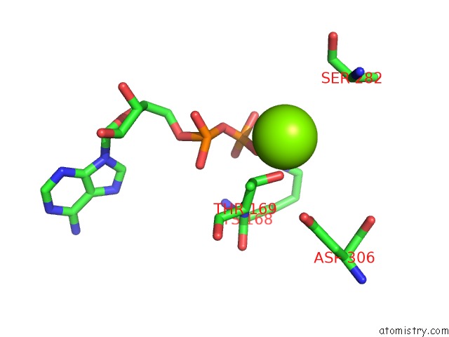
Mono view
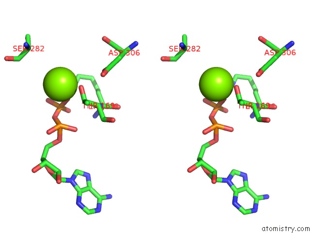
Stereo pair view
Mono view
Stereo pair view
A full contact list of Magnesium with other atoms in the Mg binding
site number 1 of The Crystal Structure of the Minimal Functional Domain of the Microtubule Destabilizer KIF2C Complexed with Mg-Adp within 5.0Å range:
|
Reference:
T.Ogawa,
R.Nitta,
Y.Okada,
N.Hirokawa.
A Common Mechanism For Microtubule Destabilizers-M Type Kinesins Stabilize Curling of the Protofilament Using the Class-Specific Neck and Loops. Cell(Cambridge,Mass.) V. 116 591 2004.
ISSN: ISSN 0092-8674
PubMed: 14980225
DOI: 10.1016/S0092-8674(04)00129-1
Page generated: Tue Aug 13 15:01:34 2024
ISSN: ISSN 0092-8674
PubMed: 14980225
DOI: 10.1016/S0092-8674(04)00129-1
Last articles
Zn in 9MJ5Zn in 9HNW
Zn in 9G0L
Zn in 9FNE
Zn in 9DZN
Zn in 9E0I
Zn in 9D32
Zn in 9DAK
Zn in 8ZXC
Zn in 8ZUF