Magnesium »
PDB 1v5g-1vq4 »
1va6 »
Magnesium in PDB 1va6: Crystal Structure of Gamma-Glutamylcysteine Synthetase From Escherichia Coli B Complexed with Transition-State Analogue
Enzymatic activity of Crystal Structure of Gamma-Glutamylcysteine Synthetase From Escherichia Coli B Complexed with Transition-State Analogue
All present enzymatic activity of Crystal Structure of Gamma-Glutamylcysteine Synthetase From Escherichia Coli B Complexed with Transition-State Analogue:
6.3.2.2;
6.3.2.2;
Protein crystallography data
The structure of Crystal Structure of Gamma-Glutamylcysteine Synthetase From Escherichia Coli B Complexed with Transition-State Analogue, PDB code: 1va6
was solved by
T.Hibi,
H.Nii,
T.Nakatsu,
H.Kato,
J.Hiratake,
J.Oda,
with X-Ray Crystallography technique. A brief refinement statistics is given in the table below:
| Resolution Low / High (Å) | 40.00 / 2.10 |
| Space group | P 1 21 1 |
| Cell size a, b, c (Å), α, β, γ (°) | 70.468, 97.360, 102.185, 90.00, 109.63, 90.00 |
| R / Rfree (%) | 20 / 22.5 |
Magnesium Binding Sites:
The binding sites of Magnesium atom in the Crystal Structure of Gamma-Glutamylcysteine Synthetase From Escherichia Coli B Complexed with Transition-State Analogue
(pdb code 1va6). This binding sites where shown within
5.0 Angstroms radius around Magnesium atom.
In total 8 binding sites of Magnesium where determined in the Crystal Structure of Gamma-Glutamylcysteine Synthetase From Escherichia Coli B Complexed with Transition-State Analogue, PDB code: 1va6:
Jump to Magnesium binding site number: 1; 2; 3; 4; 5; 6; 7; 8;
In total 8 binding sites of Magnesium where determined in the Crystal Structure of Gamma-Glutamylcysteine Synthetase From Escherichia Coli B Complexed with Transition-State Analogue, PDB code: 1va6:
Jump to Magnesium binding site number: 1; 2; 3; 4; 5; 6; 7; 8;
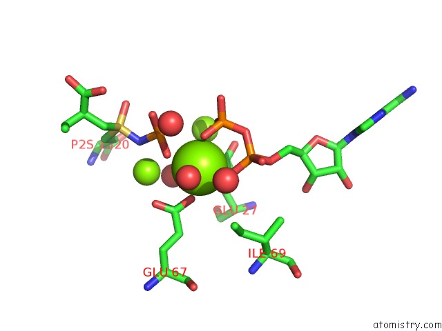
Magnesium binding site 1 out of 8 in 1va6
Go back to
Magnesium binding site 1 out
of 8 in the Crystal Structure of Gamma-Glutamylcysteine Synthetase From Escherichia Coli B Complexed with Transition-State Analogue
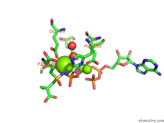
Mono view

Stereo pair view
Mono view
Stereo pair view
A full contact list of Magnesium with other atoms in the Mg binding
site number 1 of Crystal Structure of Gamma-Glutamylcysteine Synthetase From Escherichia Coli B Complexed with Transition-State Analogue within 5.0Å range:
|
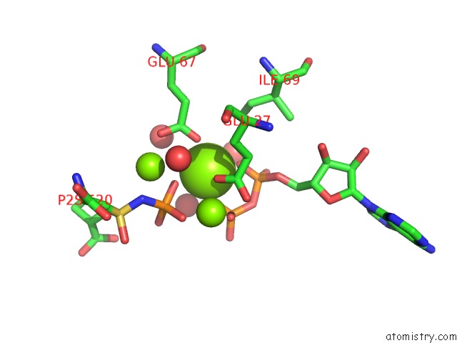
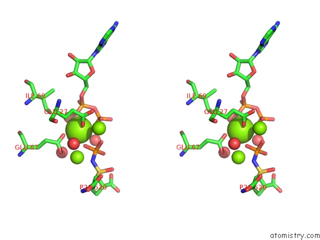
Magnesium binding site 2 out of 8 in 1va6
Go back to
Magnesium binding site 2 out
of 8 in the Crystal Structure of Gamma-Glutamylcysteine Synthetase From Escherichia Coli B Complexed with Transition-State Analogue

Mono view

Stereo pair view
Mono view
Stereo pair view
A full contact list of Magnesium with other atoms in the Mg binding
site number 2 of Crystal Structure of Gamma-Glutamylcysteine Synthetase From Escherichia Coli B Complexed with Transition-State Analogue within 5.0Å range:
|
Magnesium binding site 3 out of 8 in 1va6
Go back to
Magnesium binding site 3 out
of 8 in the Crystal Structure of Gamma-Glutamylcysteine Synthetase From Escherichia Coli B Complexed with Transition-State Analogue
Mono view
Stereo pair view
Mono view
Stereo pair view
A full contact list of Magnesium with other atoms in the Mg binding
site number 3 of Crystal Structure of Gamma-Glutamylcysteine Synthetase From Escherichia Coli B Complexed with Transition-State Analogue within 5.0Å range:
|
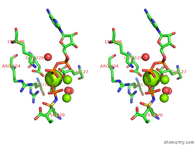
Magnesium binding site 4 out of 8 in 1va6
Go back to
Magnesium binding site 4 out
of 8 in the Crystal Structure of Gamma-Glutamylcysteine Synthetase From Escherichia Coli B Complexed with Transition-State Analogue

Mono view

Stereo pair view
Mono view
Stereo pair view
A full contact list of Magnesium with other atoms in the Mg binding
site number 4 of Crystal Structure of Gamma-Glutamylcysteine Synthetase From Escherichia Coli B Complexed with Transition-State Analogue within 5.0Å range:
|
Magnesium binding site 5 out of 8 in 1va6
Go back to
Magnesium binding site 5 out
of 8 in the Crystal Structure of Gamma-Glutamylcysteine Synthetase From Escherichia Coli B Complexed with Transition-State Analogue

Mono view

Stereo pair view
Mono view
Stereo pair view
A full contact list of Magnesium with other atoms in the Mg binding
site number 5 of Crystal Structure of Gamma-Glutamylcysteine Synthetase From Escherichia Coli B Complexed with Transition-State Analogue within 5.0Å range:
|
Magnesium binding site 6 out of 8 in 1va6
Go back to
Magnesium binding site 6 out
of 8 in the Crystal Structure of Gamma-Glutamylcysteine Synthetase From Escherichia Coli B Complexed with Transition-State Analogue

Mono view
Stereo pair view
Mono view
Stereo pair view
A full contact list of Magnesium with other atoms in the Mg binding
site number 6 of Crystal Structure of Gamma-Glutamylcysteine Synthetase From Escherichia Coli B Complexed with Transition-State Analogue within 5.0Å range:
|
Magnesium binding site 7 out of 8 in 1va6
Go back to
Magnesium binding site 7 out
of 8 in the Crystal Structure of Gamma-Glutamylcysteine Synthetase From Escherichia Coli B Complexed with Transition-State Analogue
Mono view

Stereo pair view
Mono view
Stereo pair view
A full contact list of Magnesium with other atoms in the Mg binding
site number 7 of Crystal Structure of Gamma-Glutamylcysteine Synthetase From Escherichia Coli B Complexed with Transition-State Analogue within 5.0Å range:
|
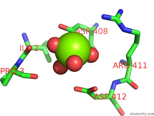
Magnesium binding site 8 out of 8 in 1va6
Go back to
Magnesium binding site 8 out
of 8 in the Crystal Structure of Gamma-Glutamylcysteine Synthetase From Escherichia Coli B Complexed with Transition-State Analogue
Mono view

Stereo pair view
Mono view
Stereo pair view
A full contact list of Magnesium with other atoms in the Mg binding
site number 8 of Crystal Structure of Gamma-Glutamylcysteine Synthetase From Escherichia Coli B Complexed with Transition-State Analogue within 5.0Å range:
|
Reference:
T.Hibi,
H.Nii,
T.Nakatsu,
A.Kimura,
H.Kato,
J.Hiratake,
J.Oda.
Crystal Structure of Gamma-Glutamylcysteine Synthetase: Insights Into the Mechanism of Catalysis By A Key Enzyme For Glutathione Homeostasis Proc.Natl.Acad.Sci.Usa V. 101 15052 2004.
ISSN: ISSN 0027-8424
PubMed: 15477603
DOI: 10.1073/PNAS.0403277101
Page generated: Tue Aug 13 15:02:02 2024
ISSN: ISSN 0027-8424
PubMed: 15477603
DOI: 10.1073/PNAS.0403277101
Last articles
Zn in 9MJ5Zn in 9HNW
Zn in 9G0L
Zn in 9FNE
Zn in 9DZN
Zn in 9E0I
Zn in 9D32
Zn in 9DAK
Zn in 8ZXC
Zn in 8ZUF