Magnesium »
PDB 1v5g-1vq4 »
1vaq »
Magnesium in PDB 1vaq: Crystal Structure of the MG2+-(Chromomycin A3)2-D(Ttggccaa)2 Complex Reveals Ggcc Binding Specificity of the Drug Dimer Chelated By Metal Ion
Protein crystallography data
The structure of Crystal Structure of the MG2+-(Chromomycin A3)2-D(Ttggccaa)2 Complex Reveals Ggcc Binding Specificity of the Drug Dimer Chelated By Metal Ion, PDB code: 1vaq
was solved by
M.H.Hou,
H.Robinson,
Y.G.Gao,
A.H.-J.Wang,
with X-Ray Crystallography technique. A brief refinement statistics is given in the table below:
| Resolution Low / High (Å) | 50.00 / 2.00 |
| Space group | P 65 2 2 |
| Cell size a, b, c (Å), α, β, γ (°) | 42.230, 42.230, 246.110, 90.00, 90.00, 120.00 |
| R / Rfree (%) | 23.5 / 27.8 |
Magnesium Binding Sites:
The binding sites of Magnesium atom in the Crystal Structure of the MG2+-(Chromomycin A3)2-D(Ttggccaa)2 Complex Reveals Ggcc Binding Specificity of the Drug Dimer Chelated By Metal Ion
(pdb code 1vaq). This binding sites where shown within
5.0 Angstroms radius around Magnesium atom.
In total 2 binding sites of Magnesium where determined in the Crystal Structure of the MG2+-(Chromomycin A3)2-D(Ttggccaa)2 Complex Reveals Ggcc Binding Specificity of the Drug Dimer Chelated By Metal Ion, PDB code: 1vaq:
Jump to Magnesium binding site number: 1; 2;
In total 2 binding sites of Magnesium where determined in the Crystal Structure of the MG2+-(Chromomycin A3)2-D(Ttggccaa)2 Complex Reveals Ggcc Binding Specificity of the Drug Dimer Chelated By Metal Ion, PDB code: 1vaq:
Jump to Magnesium binding site number: 1; 2;
Magnesium binding site 1 out of 2 in 1vaq
Go back to
Magnesium binding site 1 out
of 2 in the Crystal Structure of the MG2+-(Chromomycin A3)2-D(Ttggccaa)2 Complex Reveals Ggcc Binding Specificity of the Drug Dimer Chelated By Metal Ion
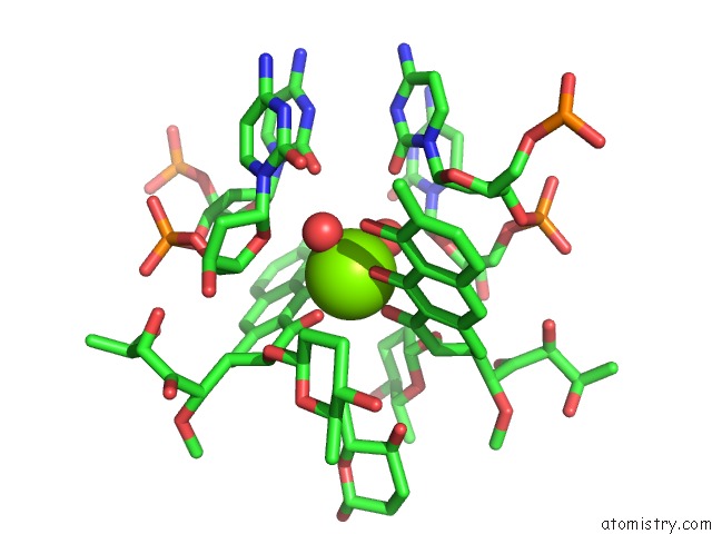
Mono view
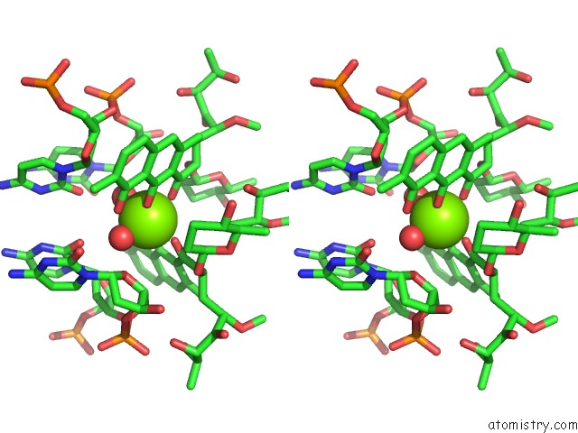
Stereo pair view
Mono view
Stereo pair view
A full contact list of Magnesium with other atoms in the Mg binding
site number 1 of Crystal Structure of the MG2+-(Chromomycin A3)2-D(Ttggccaa)2 Complex Reveals Ggcc Binding Specificity of the Drug Dimer Chelated By Metal Ion within 5.0Å range:
|
Magnesium binding site 2 out of 2 in 1vaq
Go back to
Magnesium binding site 2 out
of 2 in the Crystal Structure of the MG2+-(Chromomycin A3)2-D(Ttggccaa)2 Complex Reveals Ggcc Binding Specificity of the Drug Dimer Chelated By Metal Ion
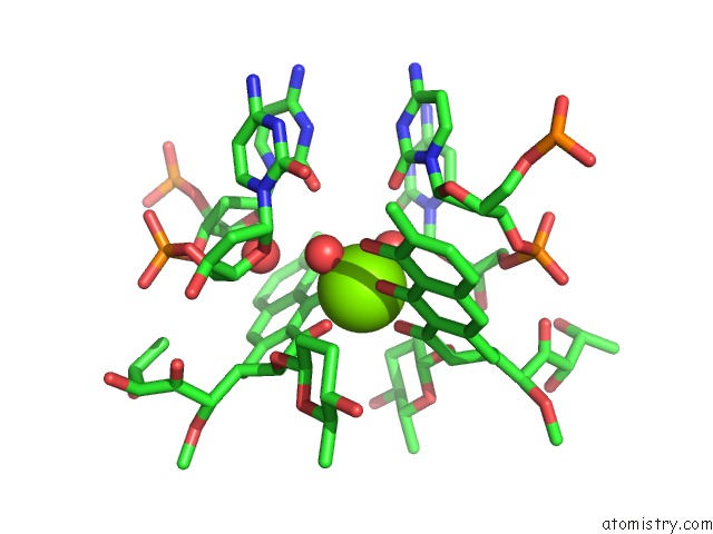
Mono view
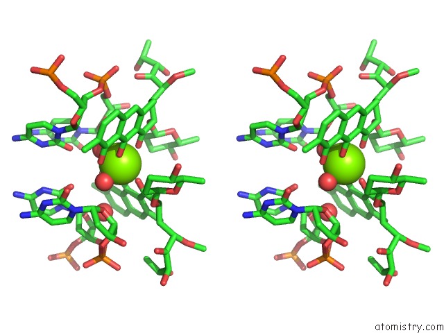
Stereo pair view
Mono view
Stereo pair view
A full contact list of Magnesium with other atoms in the Mg binding
site number 2 of Crystal Structure of the MG2+-(Chromomycin A3)2-D(Ttggccaa)2 Complex Reveals Ggcc Binding Specificity of the Drug Dimer Chelated By Metal Ion within 5.0Å range:
|
Reference:
M.H.Hou,
H.Robinson,
Y.G.Gao,
A.H.-J.Wang.
Crystal Structure of the [MG2+-(Chromomycin A3)2]-D(Ttggccaa)2 Complex Reveals Ggcc Binding Specificity of the Drug Dimer Chelated By A Metal Ion Nucleic Acids Res. V. 32 2214 2004.
ISSN: ISSN 0305-1048
PubMed: 15107489
DOI: 10.1093/NAR/GKH549
Page generated: Tue Aug 13 15:02:03 2024
ISSN: ISSN 0305-1048
PubMed: 15107489
DOI: 10.1093/NAR/GKH549
Last articles
K in 8RMHK in 8K1E
K in 7G7O
K in 7G7S
K in 7G7P
K in 7G7X
K in 7G7L
K in 7G7T
K in 7G7Q
K in 7G7R