Magnesium »
PDB 1v5g-1vq4 »
1vg9 »
Magnesium in PDB 1vg9: The Crystal Structures of the Rep-1 Protein in Complex with C- Terminally Truncated RAB7 Protein
Protein crystallography data
The structure of The Crystal Structures of the Rep-1 Protein in Complex with C- Terminally Truncated RAB7 Protein, PDB code: 1vg9
was solved by
A.Rak,
O.Pylypenko,
A.Niculae,
K.Pyatkov,
R.S.Goody,
K.Alexandrov,
with X-Ray Crystallography technique. A brief refinement statistics is given in the table below:
| Resolution Low / High (Å) | 19.42 / 2.50 |
| Space group | P 1 21 1 |
| Cell size a, b, c (Å), α, β, γ (°) | 66.392, 144.691, 200.086, 90.00, 90.13, 90.00 |
| R / Rfree (%) | 20.6 / 25 |
Other elements in 1vg9:
The structure of The Crystal Structures of the Rep-1 Protein in Complex with C- Terminally Truncated RAB7 Protein also contains other interesting chemical elements:
| Potassium | (K) | 4 atoms |
Magnesium Binding Sites:
The binding sites of Magnesium atom in the The Crystal Structures of the Rep-1 Protein in Complex with C- Terminally Truncated RAB7 Protein
(pdb code 1vg9). This binding sites where shown within
5.0 Angstroms radius around Magnesium atom.
In total 4 binding sites of Magnesium where determined in the The Crystal Structures of the Rep-1 Protein in Complex with C- Terminally Truncated RAB7 Protein, PDB code: 1vg9:
Jump to Magnesium binding site number: 1; 2; 3; 4;
In total 4 binding sites of Magnesium where determined in the The Crystal Structures of the Rep-1 Protein in Complex with C- Terminally Truncated RAB7 Protein, PDB code: 1vg9:
Jump to Magnesium binding site number: 1; 2; 3; 4;
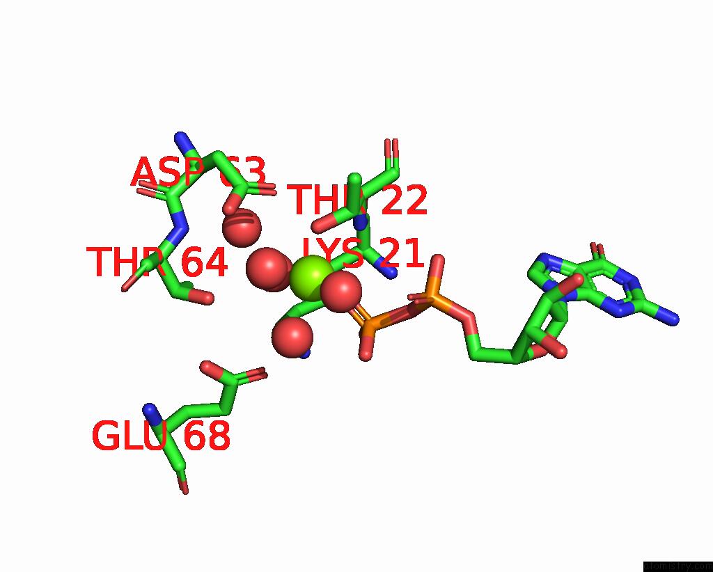
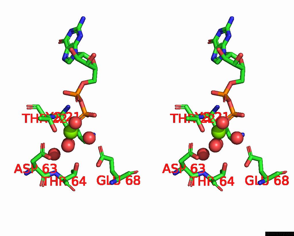
Magnesium binding site 1 out of 4 in 1vg9
Go back to
Magnesium binding site 1 out
of 4 in the The Crystal Structures of the Rep-1 Protein in Complex with C- Terminally Truncated RAB7 Protein
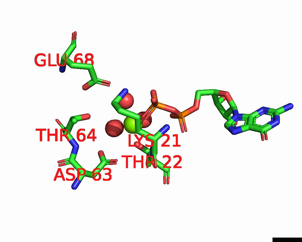
Mono view
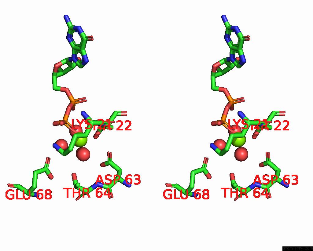
Stereo pair view
Mono view
Stereo pair view
A full contact list of Magnesium with other atoms in the Mg binding
site number 1 of The Crystal Structures of the Rep-1 Protein in Complex with C- Terminally Truncated RAB7 Protein within 5.0Å range:
|
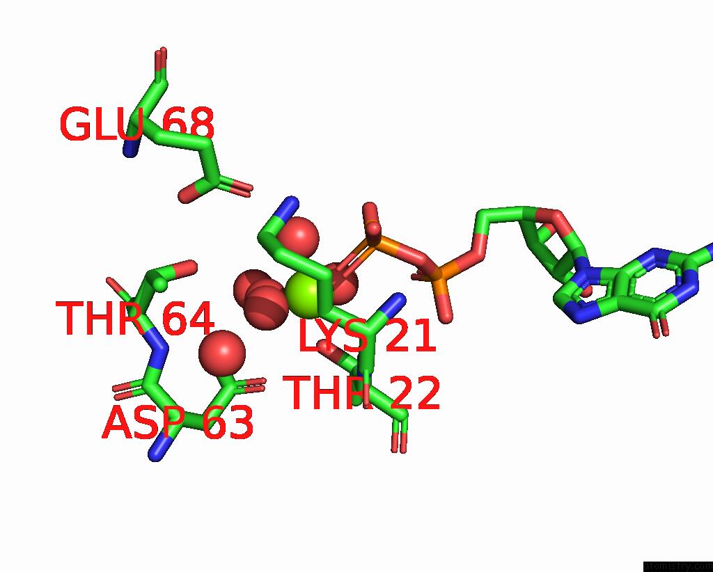
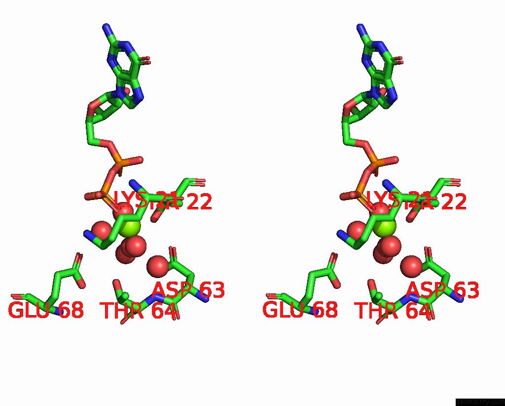
Magnesium binding site 2 out of 4 in 1vg9
Go back to
Magnesium binding site 2 out
of 4 in the The Crystal Structures of the Rep-1 Protein in Complex with C- Terminally Truncated RAB7 Protein
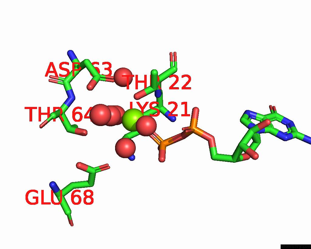
Mono view
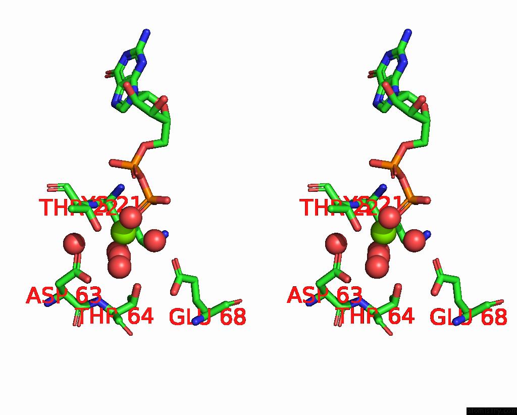
Stereo pair view
Mono view
Stereo pair view
A full contact list of Magnesium with other atoms in the Mg binding
site number 2 of The Crystal Structures of the Rep-1 Protein in Complex with C- Terminally Truncated RAB7 Protein within 5.0Å range:
|
Magnesium binding site 3 out of 4 in 1vg9
Go back to
Magnesium binding site 3 out
of 4 in the The Crystal Structures of the Rep-1 Protein in Complex with C- Terminally Truncated RAB7 Protein
Mono view
Stereo pair view
Mono view
Stereo pair view
A full contact list of Magnesium with other atoms in the Mg binding
site number 3 of The Crystal Structures of the Rep-1 Protein in Complex with C- Terminally Truncated RAB7 Protein within 5.0Å range:
|
Magnesium binding site 4 out of 4 in 1vg9
Go back to
Magnesium binding site 4 out
of 4 in the The Crystal Structures of the Rep-1 Protein in Complex with C- Terminally Truncated RAB7 Protein
Mono view
Stereo pair view
Mono view
Stereo pair view
A full contact list of Magnesium with other atoms in the Mg binding
site number 4 of The Crystal Structures of the Rep-1 Protein in Complex with C- Terminally Truncated RAB7 Protein within 5.0Å range:
|
Reference:
A.Rak,
O.Pylypenko,
A.Niculae,
K.Pyatkov,
R.S.Goody,
K.Alexandrov.
Structure of the RAB7:Rep-1 Complex: Insights Into the Mechanism of Rab Prenylation and Choroideremia Disease Cell(Cambridge,Mass.) V. 117 749 2004.
ISSN: ISSN 0092-8674
PubMed: 15186776
DOI: 10.1016/J.CELL.2004.05.017
Page generated: Tue Aug 13 15:04:42 2024
ISSN: ISSN 0092-8674
PubMed: 15186776
DOI: 10.1016/J.CELL.2004.05.017
Last articles
Zn in 9MJ5Zn in 9HNW
Zn in 9G0L
Zn in 9FNE
Zn in 9DZN
Zn in 9E0I
Zn in 9D32
Zn in 9DAK
Zn in 8ZXC
Zn in 8ZUF