Magnesium »
PDB 1v5g-1vq4 »
1vid »
Magnesium in PDB 1vid: Catechol O-Methyltransferase
Enzymatic activity of Catechol O-Methyltransferase
All present enzymatic activity of Catechol O-Methyltransferase:
2.1.1.6;
2.1.1.6;
Protein crystallography data
The structure of Catechol O-Methyltransferase, PDB code: 1vid
was solved by
J.Vidgren,
L.A.Svensson,
A.Liljas,
with X-Ray Crystallography technique. A brief refinement statistics is given in the table below:
| Resolution Low / High (Å) | 8.00 / 2.00 |
| Space group | P 32 2 1 |
| Cell size a, b, c (Å), α, β, γ (°) | 51.300, 51.300, 168.500, 90.00, 90.00, 120.00 |
| R / Rfree (%) | 19.4 / n/a |
Magnesium Binding Sites:
The binding sites of Magnesium atom in the Catechol O-Methyltransferase
(pdb code 1vid). This binding sites where shown within
5.0 Angstroms radius around Magnesium atom.
In total only one binding site of Magnesium was determined in the Catechol O-Methyltransferase, PDB code: 1vid:
In total only one binding site of Magnesium was determined in the Catechol O-Methyltransferase, PDB code: 1vid:
Magnesium binding site 1 out of 1 in 1vid
Go back to
Magnesium binding site 1 out
of 1 in the Catechol O-Methyltransferase
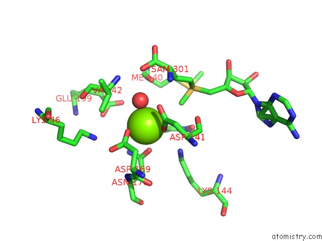
Mono view
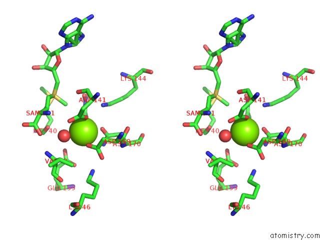
Stereo pair view
Mono view
Stereo pair view
A full contact list of Magnesium with other atoms in the Mg binding
site number 1 of Catechol O-Methyltransferase within 5.0Å range:
|
Reference:
J.Vidgren,
L.A.Svensson,
A.Liljas.
Crystal Structure of Catechol O-Methyltransferase. Nature V. 368 354 1994.
ISSN: ISSN 0028-0836
PubMed: 8127373
DOI: 10.1038/368354A0
Page generated: Tue Aug 13 15:04:41 2024
ISSN: ISSN 0028-0836
PubMed: 8127373
DOI: 10.1038/368354A0
Last articles
Ca in 5VT8Ca in 5VYF
Ca in 5VYB
Ca in 5VXZ
Ca in 5VTM
Ca in 5VWM
Ca in 5VTD
Ca in 5VUG
Ca in 5VS1
Ca in 5VRB