Magnesium »
PDB 1v5g-1vq4 »
1vma »
Magnesium in PDB 1vma: Crystal Structure of Cell Division Protein Ftsy (TM0570) From Thermotoga Maritima at 1.60 A Resolution
Protein crystallography data
The structure of Crystal Structure of Cell Division Protein Ftsy (TM0570) From Thermotoga Maritima at 1.60 A Resolution, PDB code: 1vma
was solved by
Joint Center For Structural Genomics (Jcsg),
with X-Ray Crystallography technique. A brief refinement statistics is given in the table below:
| Resolution Low / High (Å) | 24.88 / 1.60 |
| Space group | P 1 21 1 |
| Cell size a, b, c (Å), α, β, γ (°) | 78.446, 48.102, 90.792, 90.00, 107.94, 90.00 |
| R / Rfree (%) | 20.7 / 25.3 |
Magnesium Binding Sites:
The binding sites of Magnesium atom in the Crystal Structure of Cell Division Protein Ftsy (TM0570) From Thermotoga Maritima at 1.60 A Resolution
(pdb code 1vma). This binding sites where shown within
5.0 Angstroms radius around Magnesium atom.
In total only one binding site of Magnesium was determined in the Crystal Structure of Cell Division Protein Ftsy (TM0570) From Thermotoga Maritima at 1.60 A Resolution, PDB code: 1vma:
In total only one binding site of Magnesium was determined in the Crystal Structure of Cell Division Protein Ftsy (TM0570) From Thermotoga Maritima at 1.60 A Resolution, PDB code: 1vma:
Magnesium binding site 1 out of 1 in 1vma
Go back to
Magnesium binding site 1 out
of 1 in the Crystal Structure of Cell Division Protein Ftsy (TM0570) From Thermotoga Maritima at 1.60 A Resolution
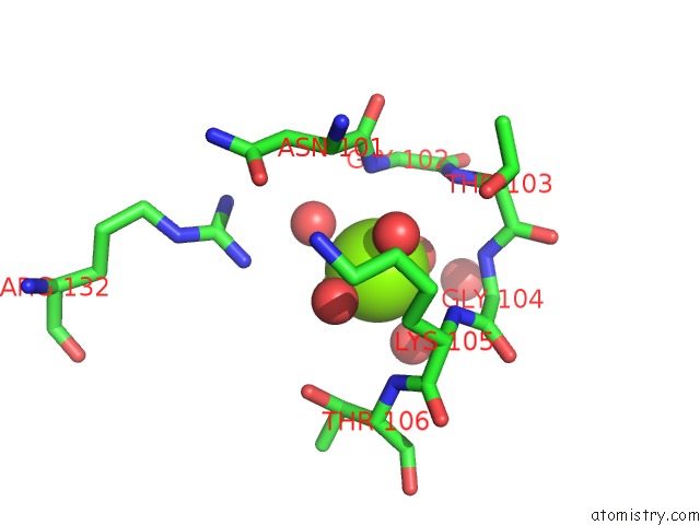
Mono view
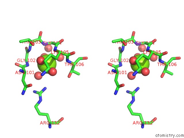
Stereo pair view
Mono view
Stereo pair view
A full contact list of Magnesium with other atoms in the Mg binding
site number 1 of Crystal Structure of Cell Division Protein Ftsy (TM0570) From Thermotoga Maritima at 1.60 A Resolution within 5.0Å range:
|
Reference:
Joint Center For Structural Genomics (Jcsg),
Joint Center For Structural Genomics (Jcsg).
N/A N/A.
Page generated: Tue Aug 13 15:06:15 2024
Last articles
Ca in 5O6NCa in 5O5U
Ca in 5NZN
Ca in 5O5T
Ca in 5O5R
Ca in 5NZF
Ca in 5O2Z
Ca in 5O2Y
Ca in 5O0Z
Ca in 5O25