Magnesium »
PDB 1vq5-1w55 »
1w2y »
Magnesium in PDB 1w2y: The Crystal Structure of A Complex of Campylobacter Jejuni Dutpase with Substrate Analogue Dupnhp
Enzymatic activity of The Crystal Structure of A Complex of Campylobacter Jejuni Dutpase with Substrate Analogue Dupnhp
All present enzymatic activity of The Crystal Structure of A Complex of Campylobacter Jejuni Dutpase with Substrate Analogue Dupnhp:
3.6.1.23;
3.6.1.23;
Protein crystallography data
The structure of The Crystal Structure of A Complex of Campylobacter Jejuni Dutpase with Substrate Analogue Dupnhp, PDB code: 1w2y
was solved by
O.V.Moroz,
M.Harkiolaki,
M.Y.Galperin,
A.A.Vagin,
D.Gonzalez-Pacanowska,
K.S.Wilson,
with X-Ray Crystallography technique. A brief refinement statistics is given in the table below:
| Resolution Low / High (Å) | 55.90 / 1.65 |
| Space group | P 21 21 21 |
| Cell size a, b, c (Å), α, β, γ (°) | 66.963, 70.629, 92.850, 90.00, 90.00, 90.00 |
| R / Rfree (%) | 15.1 / 19.4 |
Magnesium Binding Sites:
The binding sites of Magnesium atom in the The Crystal Structure of A Complex of Campylobacter Jejuni Dutpase with Substrate Analogue Dupnhp
(pdb code 1w2y). This binding sites where shown within
5.0 Angstroms radius around Magnesium atom.
In total 6 binding sites of Magnesium where determined in the The Crystal Structure of A Complex of Campylobacter Jejuni Dutpase with Substrate Analogue Dupnhp, PDB code: 1w2y:
Jump to Magnesium binding site number: 1; 2; 3; 4; 5; 6;
In total 6 binding sites of Magnesium where determined in the The Crystal Structure of A Complex of Campylobacter Jejuni Dutpase with Substrate Analogue Dupnhp, PDB code: 1w2y:
Jump to Magnesium binding site number: 1; 2; 3; 4; 5; 6;
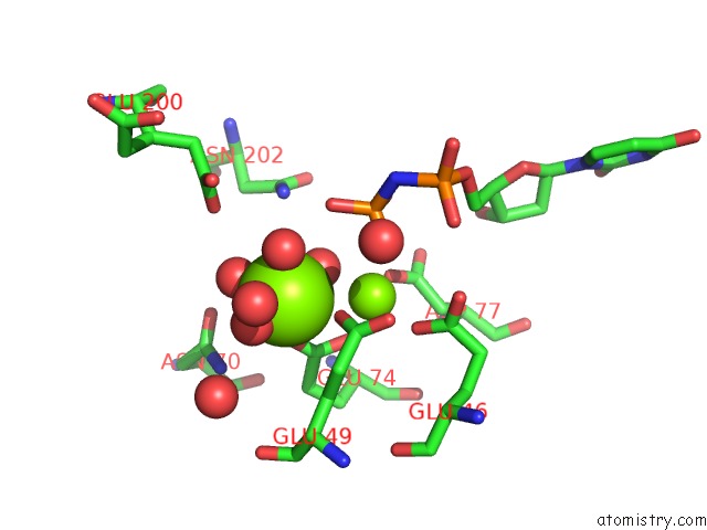
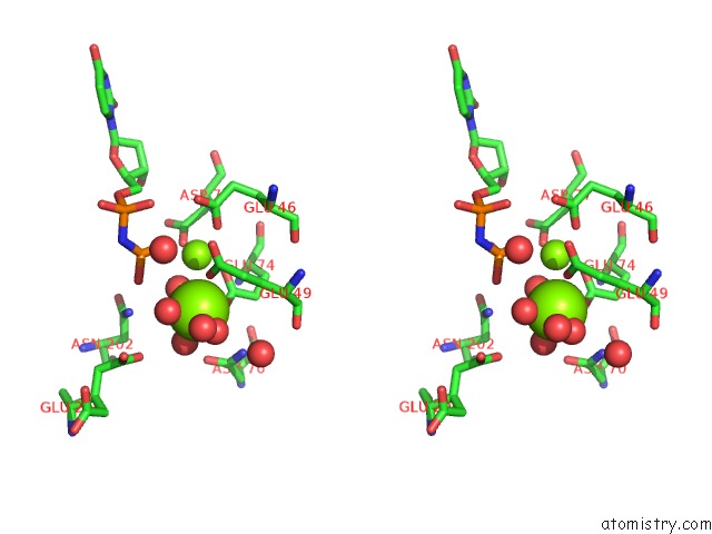
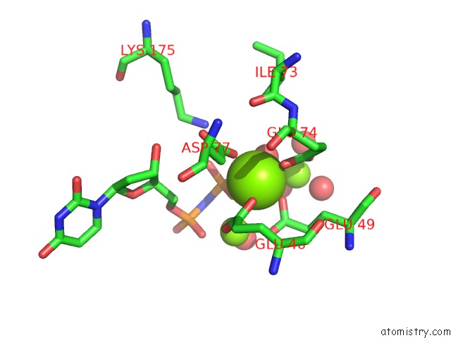
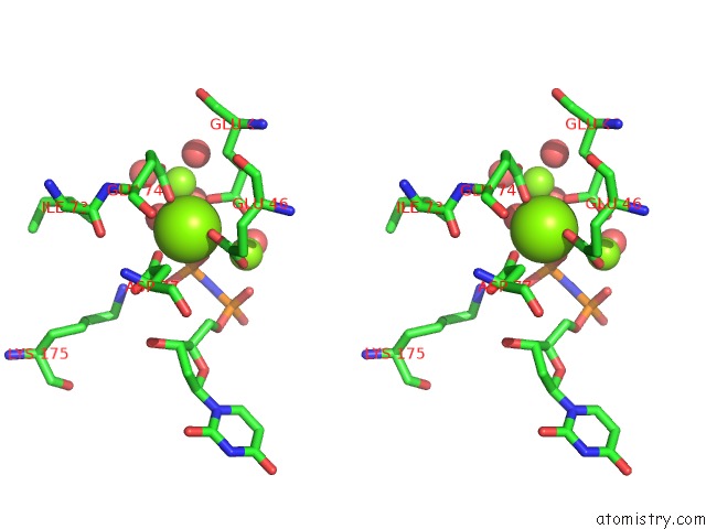
Magnesium binding site 1 out of 6 in 1w2y
Go back to
Magnesium binding site 1 out
of 6 in the The Crystal Structure of A Complex of Campylobacter Jejuni Dutpase with Substrate Analogue Dupnhp
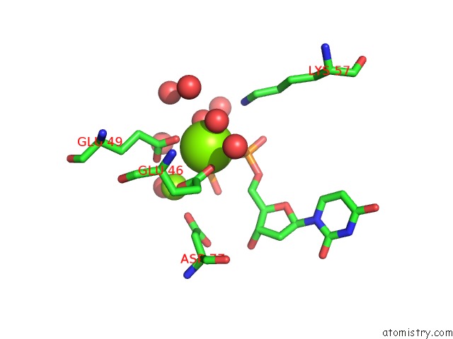
Mono view
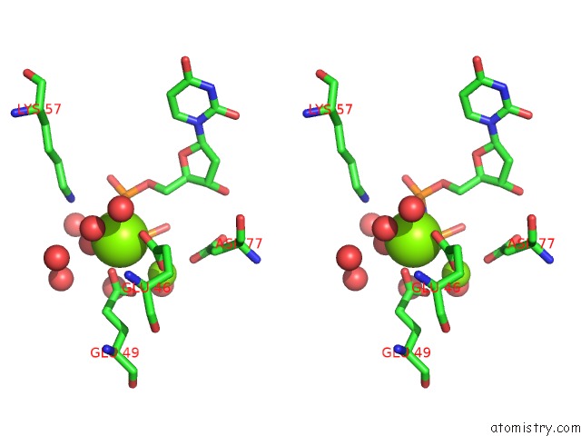
Stereo pair view
Mono view
Stereo pair view
A full contact list of Magnesium with other atoms in the Mg binding
site number 1 of The Crystal Structure of A Complex of Campylobacter Jejuni Dutpase with Substrate Analogue Dupnhp within 5.0Å range:
|
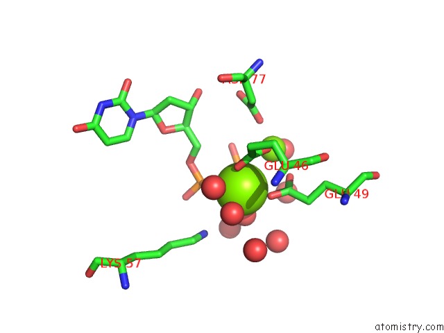
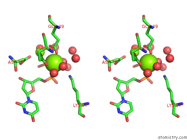
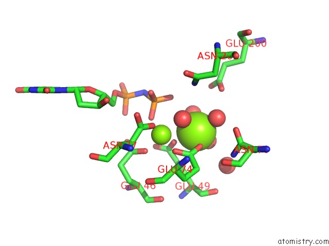
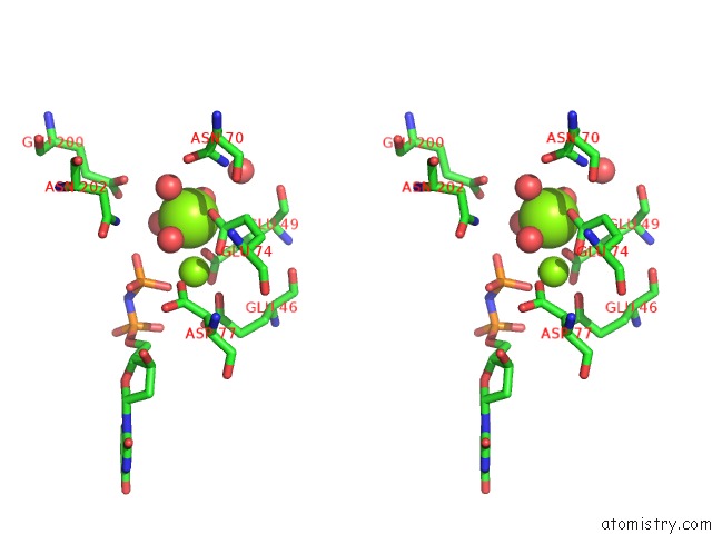
Magnesium binding site 2 out of 6 in 1w2y
Go back to
Magnesium binding site 2 out
of 6 in the The Crystal Structure of A Complex of Campylobacter Jejuni Dutpase with Substrate Analogue Dupnhp
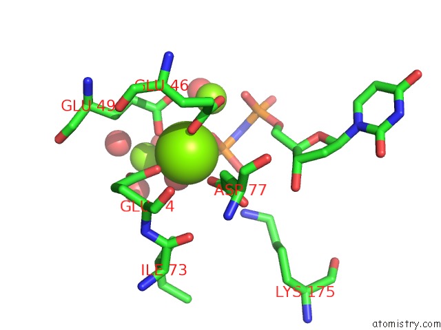
Mono view
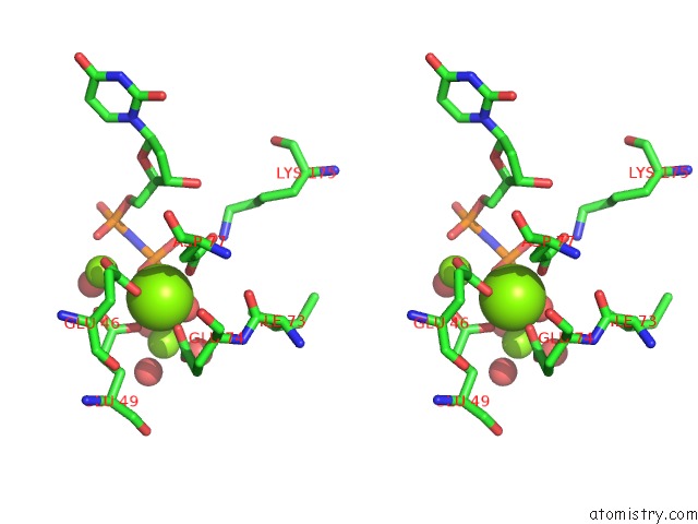
Stereo pair view
Mono view
Stereo pair view
A full contact list of Magnesium with other atoms in the Mg binding
site number 2 of The Crystal Structure of A Complex of Campylobacter Jejuni Dutpase with Substrate Analogue Dupnhp within 5.0Å range:
|
Magnesium binding site 3 out of 6 in 1w2y
Go back to
Magnesium binding site 3 out
of 6 in the The Crystal Structure of A Complex of Campylobacter Jejuni Dutpase with Substrate Analogue Dupnhp
Mono view
Stereo pair view
Mono view
Stereo pair view
A full contact list of Magnesium with other atoms in the Mg binding
site number 3 of The Crystal Structure of A Complex of Campylobacter Jejuni Dutpase with Substrate Analogue Dupnhp within 5.0Å range:
|
Magnesium binding site 4 out of 6 in 1w2y
Go back to
Magnesium binding site 4 out
of 6 in the The Crystal Structure of A Complex of Campylobacter Jejuni Dutpase with Substrate Analogue Dupnhp
Mono view
Stereo pair view
Mono view
Stereo pair view
A full contact list of Magnesium with other atoms in the Mg binding
site number 4 of The Crystal Structure of A Complex of Campylobacter Jejuni Dutpase with Substrate Analogue Dupnhp within 5.0Å range:
|
Magnesium binding site 5 out of 6 in 1w2y
Go back to
Magnesium binding site 5 out
of 6 in the The Crystal Structure of A Complex of Campylobacter Jejuni Dutpase with Substrate Analogue Dupnhp
Mono view
Stereo pair view
Mono view
Stereo pair view
A full contact list of Magnesium with other atoms in the Mg binding
site number 5 of The Crystal Structure of A Complex of Campylobacter Jejuni Dutpase with Substrate Analogue Dupnhp within 5.0Å range:
|
Magnesium binding site 6 out of 6 in 1w2y
Go back to
Magnesium binding site 6 out
of 6 in the The Crystal Structure of A Complex of Campylobacter Jejuni Dutpase with Substrate Analogue Dupnhp
Mono view
Stereo pair view
Mono view
Stereo pair view
A full contact list of Magnesium with other atoms in the Mg binding
site number 6 of The Crystal Structure of A Complex of Campylobacter Jejuni Dutpase with Substrate Analogue Dupnhp within 5.0Å range:
|
Reference:
O.V.Moroz,
M.Harkiolaki,
M.Y.Galperin,
A.A.Vagin,
D.Gonzalez-Pacanowska,
K.S.Wilson.
The Crystal Structure of A Complex of Campylobacter Jejuni Dutpase with Substrate Analogue Sheds Light on the Mechanism and Suggests the "Basic Module" For Dimeric D(C/U)Tpases J.Mol.Biol. V. 342 1583 2004.
ISSN: ISSN 0022-2836
PubMed: 15364583
DOI: 10.1016/J.JMB.2004.07.050
Page generated: Sun Aug 10 06:31:27 2025
ISSN: ISSN 0022-2836
PubMed: 15364583
DOI: 10.1016/J.JMB.2004.07.050
Last articles
Mg in 2M2WMg in 2M2U
Mg in 2M32
Mg in 2LWI
Mg in 2LVJ
Mg in 2L4I
Mg in 2LCF
Mg in 2KWI
Mg in 2L0X
Mg in 2KSZ