Magnesium »
PDB 1w56-1wdt »
1w6t »
Magnesium in PDB 1w6t: Crystal Structure of Octameric Enolase From Streptococcus Pneumoniae
Enzymatic activity of Crystal Structure of Octameric Enolase From Streptococcus Pneumoniae
All present enzymatic activity of Crystal Structure of Octameric Enolase From Streptococcus Pneumoniae:
4.2.1.11;
4.2.1.11;
Protein crystallography data
The structure of Crystal Structure of Octameric Enolase From Streptococcus Pneumoniae, PDB code: 1w6t
was solved by
S.Ehinger,
W.-D.Schubert,
S.Bergmann,
S.Hammerschmidt,
D.W.Heinz,
with X-Ray Crystallography technique. A brief refinement statistics is given in the table below:
| Resolution Low / High (Å) | 100.00 / 2.10 |
| Space group | I 4 |
| Cell size a, b, c (Å), α, β, γ (°) | 143.702, 143.702, 100.579, 90.00, 90.00, 90.00 |
| R / Rfree (%) | 14.8 / 18.7 |
Magnesium Binding Sites:
The binding sites of Magnesium atom in the Crystal Structure of Octameric Enolase From Streptococcus Pneumoniae
(pdb code 1w6t). This binding sites where shown within
5.0 Angstroms radius around Magnesium atom.
In total 2 binding sites of Magnesium where determined in the Crystal Structure of Octameric Enolase From Streptococcus Pneumoniae, PDB code: 1w6t:
Jump to Magnesium binding site number: 1; 2;
In total 2 binding sites of Magnesium where determined in the Crystal Structure of Octameric Enolase From Streptococcus Pneumoniae, PDB code: 1w6t:
Jump to Magnesium binding site number: 1; 2;
Magnesium binding site 1 out of 2 in 1w6t
Go back to
Magnesium binding site 1 out
of 2 in the Crystal Structure of Octameric Enolase From Streptococcus Pneumoniae
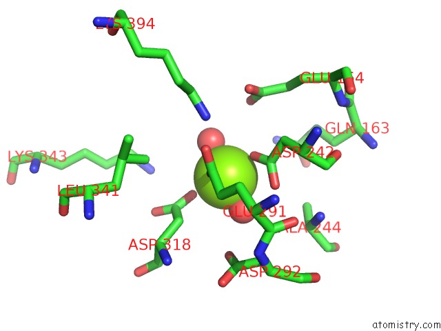
Mono view
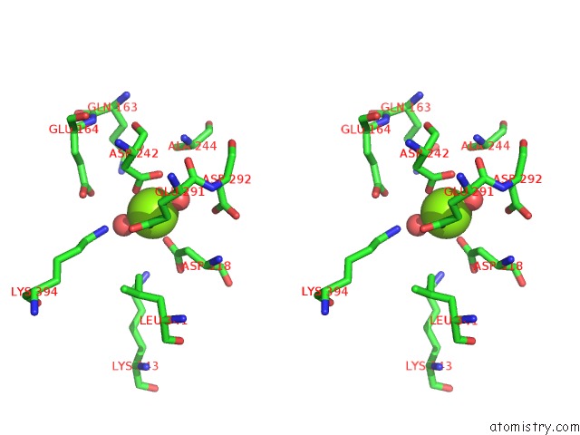
Stereo pair view
Mono view
Stereo pair view
A full contact list of Magnesium with other atoms in the Mg binding
site number 1 of Crystal Structure of Octameric Enolase From Streptococcus Pneumoniae within 5.0Å range:
|
Magnesium binding site 2 out of 2 in 1w6t
Go back to
Magnesium binding site 2 out
of 2 in the Crystal Structure of Octameric Enolase From Streptococcus Pneumoniae
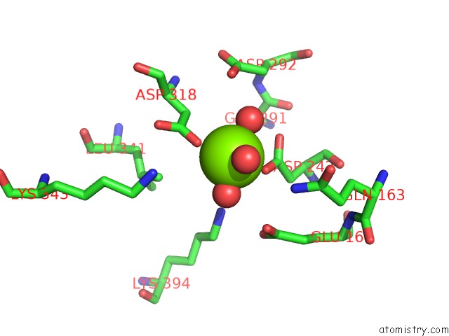
Mono view

Stereo pair view
Mono view
Stereo pair view
A full contact list of Magnesium with other atoms in the Mg binding
site number 2 of Crystal Structure of Octameric Enolase From Streptococcus Pneumoniae within 5.0Å range:
|
Reference:
S.Ehinger,
W.-D.Schubert,
S.Bergmann,
S.Hammerschmidt,
D.W.Heinz.
Plasmin(Ogen)-Binding Alpha-Enolase From Streptococcus Pneumoniae: Crystal Structure and Evaluation of Plasmin(Ogen)-Binding Sites J.Mol.Biol. V. 343 997 2004.
ISSN: ISSN 0022-2836
PubMed: 15476816
DOI: 10.1016/J.JMB.2004.08.088
Page generated: Sun Aug 10 06:43:51 2025
ISSN: ISSN 0022-2836
PubMed: 15476816
DOI: 10.1016/J.JMB.2004.08.088
Last articles
Mg in 2AETMg in 2AER
Mg in 2AEL
Mg in 2AEK
Mg in 2AE6
Mg in 2AD5
Mg in 2ACX
Mg in 2A9R
Mg in 2A9K
Mg in 2A9F