Magnesium »
PDB 1w56-1wdt »
1w8y »
Magnesium in PDB 1w8y: Crystal Structure of the Nitrocefin Acyl-Dd-Peptidase From Actinomadura R39.
Enzymatic activity of Crystal Structure of the Nitrocefin Acyl-Dd-Peptidase From Actinomadura R39.
All present enzymatic activity of Crystal Structure of the Nitrocefin Acyl-Dd-Peptidase From Actinomadura R39.:
3.4.16.4;
3.4.16.4;
Protein crystallography data
The structure of Crystal Structure of the Nitrocefin Acyl-Dd-Peptidase From Actinomadura R39., PDB code: 1w8y
was solved by
E.Sauvage,
R.Herman,
S.Petrella,
C.Duez,
J.M.Frere,
P.Charlier,
with X-Ray Crystallography technique. A brief refinement statistics is given in the table below:
| Resolution Low / High (Å) | 19.94 / 2.40 |
| Space group | P 1 21 1 |
| Cell size a, b, c (Å), α, β, γ (°) | 103.490, 94.360, 107.200, 90.00, 94.58, 90.00 |
| R / Rfree (%) | 22 / 27.7 |
Magnesium Binding Sites:
The binding sites of Magnesium atom in the Crystal Structure of the Nitrocefin Acyl-Dd-Peptidase From Actinomadura R39.
(pdb code 1w8y). This binding sites where shown within
5.0 Angstroms radius around Magnesium atom.
In total 4 binding sites of Magnesium where determined in the Crystal Structure of the Nitrocefin Acyl-Dd-Peptidase From Actinomadura R39., PDB code: 1w8y:
Jump to Magnesium binding site number: 1; 2; 3; 4;
In total 4 binding sites of Magnesium where determined in the Crystal Structure of the Nitrocefin Acyl-Dd-Peptidase From Actinomadura R39., PDB code: 1w8y:
Jump to Magnesium binding site number: 1; 2; 3; 4;
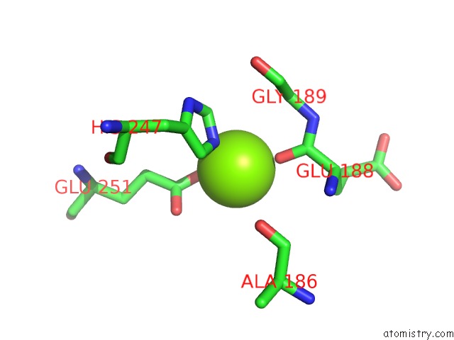
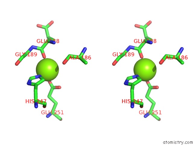
Magnesium binding site 1 out of 4 in 1w8y
Go back to
Magnesium binding site 1 out
of 4 in the Crystal Structure of the Nitrocefin Acyl-Dd-Peptidase From Actinomadura R39.
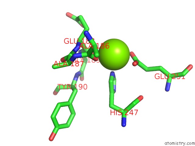
Mono view
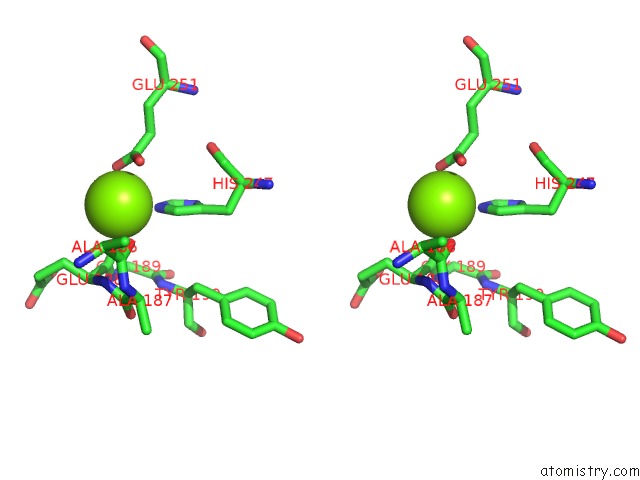
Stereo pair view
Mono view
Stereo pair view
A full contact list of Magnesium with other atoms in the Mg binding
site number 1 of Crystal Structure of the Nitrocefin Acyl-Dd-Peptidase From Actinomadura R39. within 5.0Å range:
|
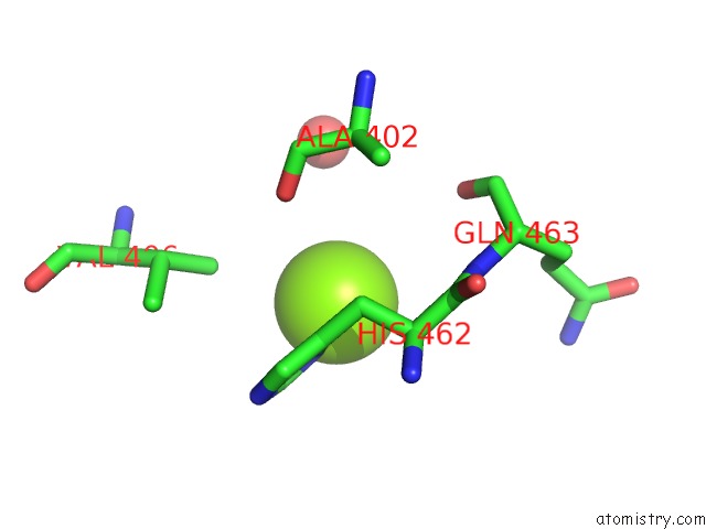
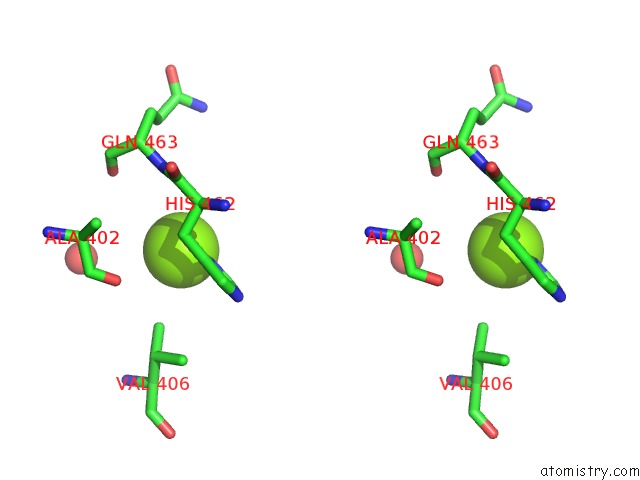
Magnesium binding site 2 out of 4 in 1w8y
Go back to
Magnesium binding site 2 out
of 4 in the Crystal Structure of the Nitrocefin Acyl-Dd-Peptidase From Actinomadura R39.
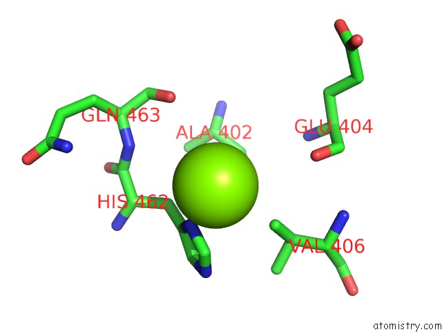
Mono view
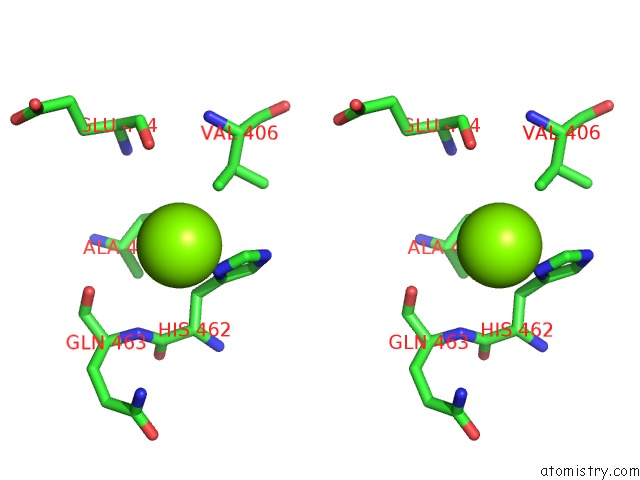
Stereo pair view
Mono view
Stereo pair view
A full contact list of Magnesium with other atoms in the Mg binding
site number 2 of Crystal Structure of the Nitrocefin Acyl-Dd-Peptidase From Actinomadura R39. within 5.0Å range:
|
Magnesium binding site 3 out of 4 in 1w8y
Go back to
Magnesium binding site 3 out
of 4 in the Crystal Structure of the Nitrocefin Acyl-Dd-Peptidase From Actinomadura R39.
Mono view
Stereo pair view
Mono view
Stereo pair view
A full contact list of Magnesium with other atoms in the Mg binding
site number 3 of Crystal Structure of the Nitrocefin Acyl-Dd-Peptidase From Actinomadura R39. within 5.0Å range:
|
Magnesium binding site 4 out of 4 in 1w8y
Go back to
Magnesium binding site 4 out
of 4 in the Crystal Structure of the Nitrocefin Acyl-Dd-Peptidase From Actinomadura R39.
Mono view
Stereo pair view
Mono view
Stereo pair view
A full contact list of Magnesium with other atoms in the Mg binding
site number 4 of Crystal Structure of the Nitrocefin Acyl-Dd-Peptidase From Actinomadura R39. within 5.0Å range:
|
Reference:
E.Sauvage,
R.Herman,
S.Petrella,
C.Duez,
F.Bouillenne,
J.M.Frere,
P.Charlier.
Crystal Structure of the Actinomadura R39 Dd- Peptidase Reveals New Domains in Penicillin- Binding Proteins. J.Biol.Chem. V. 280 31249 2005.
ISSN: ISSN 0021-9258
PubMed: 15987687
DOI: 10.1074/JBC.M503271200
Page generated: Sun Aug 10 06:46:18 2025
ISSN: ISSN 0021-9258
PubMed: 15987687
DOI: 10.1074/JBC.M503271200
Last articles
Mg in 2PU3Mg in 2PS5
Mg in 2PS7
Mg in 2PS2
Mg in 2PS4
Mg in 2PRC
Mg in 2PS1
Mg in 2PRY
Mg in 2PRN
Mg in 2PPQ