Magnesium »
PDB 1we2-1x1s »
1wzc »
Magnesium in PDB 1wzc: Crystal Structure of Pyrococcus Horikoshii Mannosyl-3- Phosphoglycerate Phosphatase Complexed with MG2+ and Phosphate
Enzymatic activity of Crystal Structure of Pyrococcus Horikoshii Mannosyl-3- Phosphoglycerate Phosphatase Complexed with MG2+ and Phosphate
All present enzymatic activity of Crystal Structure of Pyrococcus Horikoshii Mannosyl-3- Phosphoglycerate Phosphatase Complexed with MG2+ and Phosphate:
3.1.3.70;
3.1.3.70;
Protein crystallography data
The structure of Crystal Structure of Pyrococcus Horikoshii Mannosyl-3- Phosphoglycerate Phosphatase Complexed with MG2+ and Phosphate, PDB code: 1wzc
was solved by
T.Kawamura,
N.Watanabe,
I.Tanaka,
with X-Ray Crystallography technique. A brief refinement statistics is given in the table below:
| Resolution Low / High (Å) | 50.00 / 1.90 |
| Space group | P 1 21 1 |
| Cell size a, b, c (Å), α, β, γ (°) | 59.833, 70.691, 67.911, 90.00, 98.15, 90.00 |
| R / Rfree (%) | 21.9 / 25.3 |
Magnesium Binding Sites:
The binding sites of Magnesium atom in the Crystal Structure of Pyrococcus Horikoshii Mannosyl-3- Phosphoglycerate Phosphatase Complexed with MG2+ and Phosphate
(pdb code 1wzc). This binding sites where shown within
5.0 Angstroms radius around Magnesium atom.
In total 2 binding sites of Magnesium where determined in the Crystal Structure of Pyrococcus Horikoshii Mannosyl-3- Phosphoglycerate Phosphatase Complexed with MG2+ and Phosphate, PDB code: 1wzc:
Jump to Magnesium binding site number: 1; 2;
In total 2 binding sites of Magnesium where determined in the Crystal Structure of Pyrococcus Horikoshii Mannosyl-3- Phosphoglycerate Phosphatase Complexed with MG2+ and Phosphate, PDB code: 1wzc:
Jump to Magnesium binding site number: 1; 2;
Magnesium binding site 1 out of 2 in 1wzc
Go back to
Magnesium binding site 1 out
of 2 in the Crystal Structure of Pyrococcus Horikoshii Mannosyl-3- Phosphoglycerate Phosphatase Complexed with MG2+ and Phosphate
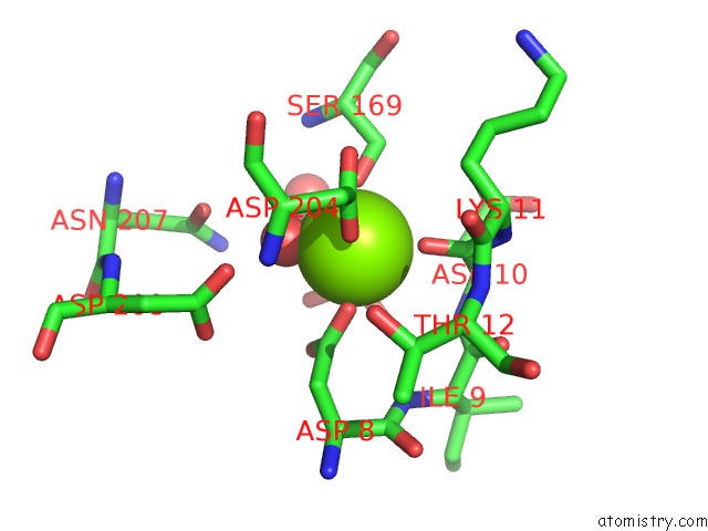
Mono view
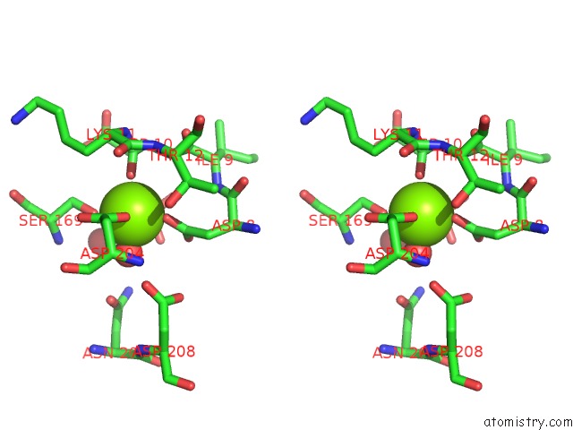
Stereo pair view
Mono view
Stereo pair view
A full contact list of Magnesium with other atoms in the Mg binding
site number 1 of Crystal Structure of Pyrococcus Horikoshii Mannosyl-3- Phosphoglycerate Phosphatase Complexed with MG2+ and Phosphate within 5.0Å range:
|
Magnesium binding site 2 out of 2 in 1wzc
Go back to
Magnesium binding site 2 out
of 2 in the Crystal Structure of Pyrococcus Horikoshii Mannosyl-3- Phosphoglycerate Phosphatase Complexed with MG2+ and Phosphate
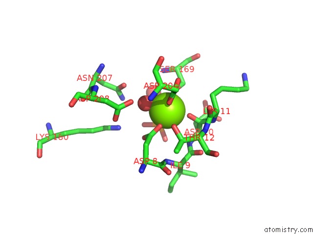
Mono view

Stereo pair view
Mono view
Stereo pair view
A full contact list of Magnesium with other atoms in the Mg binding
site number 2 of Crystal Structure of Pyrococcus Horikoshii Mannosyl-3- Phosphoglycerate Phosphatase Complexed with MG2+ and Phosphate within 5.0Å range:
|
Reference:
T.Kawamura,
N.Watanabe,
I.Tanaka.
Structure of Mannosyl-3-Phosphoglycerate Phosphatase From Pyrococcus Horikoshii. Acta Crystallogr.,Sect.D V. 64 1267 2008.
ISSN: ISSN 0907-4449
PubMed: 19018103
DOI: 10.1107/S0907444908033817
Page generated: Tue Aug 13 17:26:35 2024
ISSN: ISSN 0907-4449
PubMed: 19018103
DOI: 10.1107/S0907444908033817
Last articles
Zn in 9MJ5Zn in 9HNW
Zn in 9G0L
Zn in 9FNE
Zn in 9DZN
Zn in 9E0I
Zn in 9D32
Zn in 9DAK
Zn in 8ZXC
Zn in 8ZUF