Magnesium »
PDB 1xfy-1xon »
1xg3 »
Magnesium in PDB 1xg3: Crystal Structure of the C123S 2-Methylisocitrate Lyase Mutant From Escherichia Coli in Complex with the Reaction Product, Mg(II)-Pyruvate and Succinate
Enzymatic activity of Crystal Structure of the C123S 2-Methylisocitrate Lyase Mutant From Escherichia Coli in Complex with the Reaction Product, Mg(II)-Pyruvate and Succinate
All present enzymatic activity of Crystal Structure of the C123S 2-Methylisocitrate Lyase Mutant From Escherichia Coli in Complex with the Reaction Product, Mg(II)-Pyruvate and Succinate:
4.1.3.30;
4.1.3.30;
Protein crystallography data
The structure of Crystal Structure of the C123S 2-Methylisocitrate Lyase Mutant From Escherichia Coli in Complex with the Reaction Product, Mg(II)-Pyruvate and Succinate, PDB code: 1xg3
was solved by
S.Liu,
Z.Lu,
Y.Han,
E.Melamud,
D.Dunaway-Mariano,
O.Herzberg,
with X-Ray Crystallography technique. A brief refinement statistics is given in the table below:
| Resolution Low / High (Å) | 25.85 / 1.90 |
| Space group | C 1 2 1 |
| Cell size a, b, c (Å), α, β, γ (°) | 159.358, 85.993, 99.597, 90.00, 108.28, 90.00 |
| R / Rfree (%) | 16.1 / 19.5 |
Magnesium Binding Sites:
The binding sites of Magnesium atom in the Crystal Structure of the C123S 2-Methylisocitrate Lyase Mutant From Escherichia Coli in Complex with the Reaction Product, Mg(II)-Pyruvate and Succinate
(pdb code 1xg3). This binding sites where shown within
5.0 Angstroms radius around Magnesium atom.
In total 4 binding sites of Magnesium where determined in the Crystal Structure of the C123S 2-Methylisocitrate Lyase Mutant From Escherichia Coli in Complex with the Reaction Product, Mg(II)-Pyruvate and Succinate, PDB code: 1xg3:
Jump to Magnesium binding site number: 1; 2; 3; 4;
In total 4 binding sites of Magnesium where determined in the Crystal Structure of the C123S 2-Methylisocitrate Lyase Mutant From Escherichia Coli in Complex with the Reaction Product, Mg(II)-Pyruvate and Succinate, PDB code: 1xg3:
Jump to Magnesium binding site number: 1; 2; 3; 4;
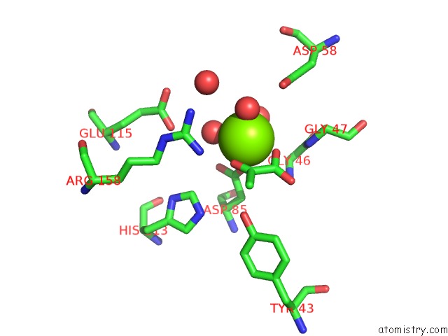
Magnesium binding site 1 out of 4 in 1xg3
Go back to
Magnesium binding site 1 out
of 4 in the Crystal Structure of the C123S 2-Methylisocitrate Lyase Mutant From Escherichia Coli in Complex with the Reaction Product, Mg(II)-Pyruvate and Succinate
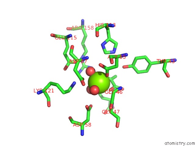
Mono view
Stereo pair view
Mono view
Stereo pair view
A full contact list of Magnesium with other atoms in the Mg binding
site number 1 of Crystal Structure of the C123S 2-Methylisocitrate Lyase Mutant From Escherichia Coli in Complex with the Reaction Product, Mg(II)-Pyruvate and Succinate within 5.0Å range:
|
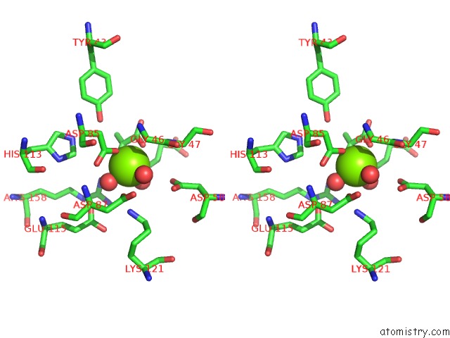
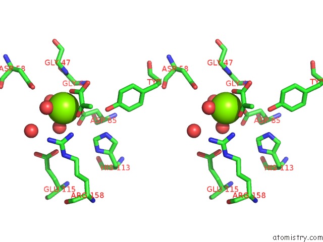
Magnesium binding site 2 out of 4 in 1xg3
Go back to
Magnesium binding site 2 out
of 4 in the Crystal Structure of the C123S 2-Methylisocitrate Lyase Mutant From Escherichia Coli in Complex with the Reaction Product, Mg(II)-Pyruvate and Succinate
Mono view
Stereo pair view
Mono view
Stereo pair view
A full contact list of Magnesium with other atoms in the Mg binding
site number 2 of Crystal Structure of the C123S 2-Methylisocitrate Lyase Mutant From Escherichia Coli in Complex with the Reaction Product, Mg(II)-Pyruvate and Succinate within 5.0Å range:
|
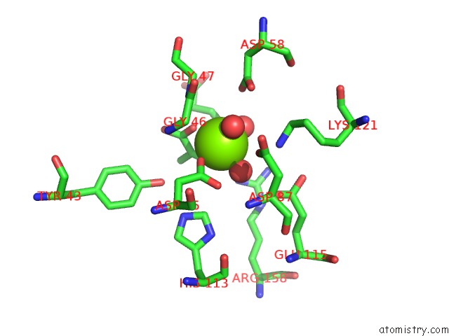
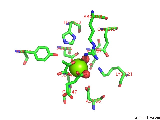
Magnesium binding site 3 out of 4 in 1xg3
Go back to
Magnesium binding site 3 out
of 4 in the Crystal Structure of the C123S 2-Methylisocitrate Lyase Mutant From Escherichia Coli in Complex with the Reaction Product, Mg(II)-Pyruvate and Succinate
Mono view
Stereo pair view
Mono view
Stereo pair view
A full contact list of Magnesium with other atoms in the Mg binding
site number 3 of Crystal Structure of the C123S 2-Methylisocitrate Lyase Mutant From Escherichia Coli in Complex with the Reaction Product, Mg(II)-Pyruvate and Succinate within 5.0Å range:
|
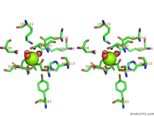
Magnesium binding site 4 out of 4 in 1xg3
Go back to
Magnesium binding site 4 out
of 4 in the Crystal Structure of the C123S 2-Methylisocitrate Lyase Mutant From Escherichia Coli in Complex with the Reaction Product, Mg(II)-Pyruvate and Succinate
Mono view
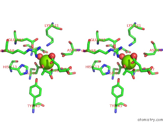
Stereo pair view
Mono view
Stereo pair view
A full contact list of Magnesium with other atoms in the Mg binding
site number 4 of Crystal Structure of the C123S 2-Methylisocitrate Lyase Mutant From Escherichia Coli in Complex with the Reaction Product, Mg(II)-Pyruvate and Succinate within 5.0Å range:
|
Reference:
S.Liu,
Z.Lu,
Y.Han,
E.Melamud,
D.Dunaway-Mariano,
O.Herzberg.
Crystal Structures of 2-Methylisocitrate Lyase in Complex with Product and with Isocitrate Inhibitor Provide Insight Into Lyase Substrate Specificity, Catalysis and Evolution Biochemistry V. 44 2949 2005.
ISSN: ISSN 0006-2960
PubMed: 15723538
DOI: 10.1021/BI0479712
Page generated: Tue Aug 13 17:39:49 2024
ISSN: ISSN 0006-2960
PubMed: 15723538
DOI: 10.1021/BI0479712
Last articles
Fe in 2YXOFe in 2YRS
Fe in 2YXC
Fe in 2YNM
Fe in 2YVJ
Fe in 2YP1
Fe in 2YU2
Fe in 2YU1
Fe in 2YQB
Fe in 2YOO