Magnesium »
PDB 1xoq-1xyc »
1xos »
Magnesium in PDB 1xos: Catalytic Domain of Human Phosphodiesterase 4B in Complex with Sildenafil
Enzymatic activity of Catalytic Domain of Human Phosphodiesterase 4B in Complex with Sildenafil
All present enzymatic activity of Catalytic Domain of Human Phosphodiesterase 4B in Complex with Sildenafil:
3.1.4.17;
3.1.4.17;
Protein crystallography data
The structure of Catalytic Domain of Human Phosphodiesterase 4B in Complex with Sildenafil, PDB code: 1xos
was solved by
G.L.Card,
B.P.England,
Y.Suzuki,
D.Fong,
B.Powell,
B.Lee,
C.Luu,
M.Tabrizizad,
S.Gillette,
P.N.Ibrahim,
D.R.Artis,
G.Bollag,
M.V.Milburn,
S.-H.Kim,
J.Schlessinger,
K.Y.J.Zhang,
with X-Ray Crystallography technique. A brief refinement statistics is given in the table below:
| Resolution Low / High (Å) | 70.71 / 2.28 |
| Space group | I 21 21 21 |
| Cell size a, b, c (Å), α, β, γ (°) | 94.571, 107.117, 89.481, 90.00, 90.00, 90.00 |
| R / Rfree (%) | 21 / 25.7 |
Other elements in 1xos:
The structure of Catalytic Domain of Human Phosphodiesterase 4B in Complex with Sildenafil also contains other interesting chemical elements:
| Zinc | (Zn) | 1 atom |
Magnesium Binding Sites:
The binding sites of Magnesium atom in the Catalytic Domain of Human Phosphodiesterase 4B in Complex with Sildenafil
(pdb code 1xos). This binding sites where shown within
5.0 Angstroms radius around Magnesium atom.
In total only one binding site of Magnesium was determined in the Catalytic Domain of Human Phosphodiesterase 4B in Complex with Sildenafil, PDB code: 1xos:
In total only one binding site of Magnesium was determined in the Catalytic Domain of Human Phosphodiesterase 4B in Complex with Sildenafil, PDB code: 1xos:
Magnesium binding site 1 out of 1 in 1xos
Go back to
Magnesium binding site 1 out
of 1 in the Catalytic Domain of Human Phosphodiesterase 4B in Complex with Sildenafil
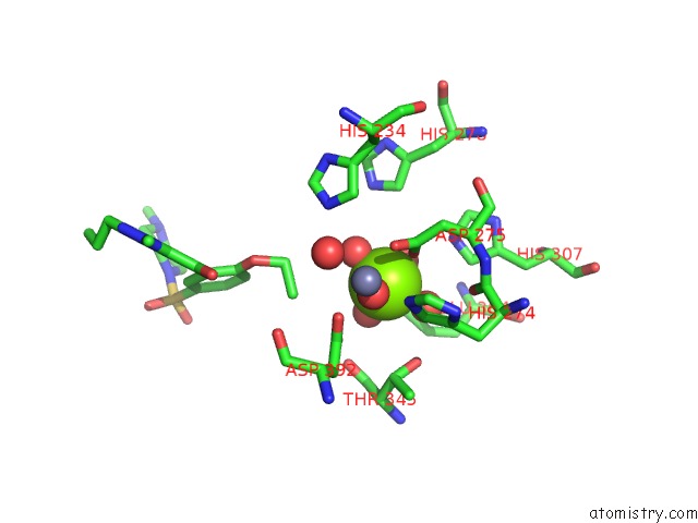
Mono view
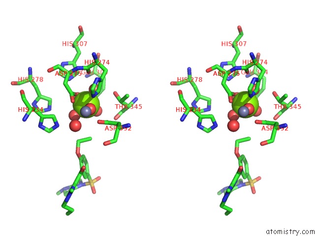
Stereo pair view
Mono view
Stereo pair view
A full contact list of Magnesium with other atoms in the Mg binding
site number 1 of Catalytic Domain of Human Phosphodiesterase 4B in Complex with Sildenafil within 5.0Å range:
|
Reference:
G.L.Card,
B.P.England,
Y.Suzuki,
D.Fong,
B.Powell,
B.Lee,
C.Luu,
M.Tabrizizad,
S.Gillette,
P.N.Ibrahim,
D.R.Artis,
G.Bollag,
M.V.Milburn,
S.-H.Kim,
J.Schlessinger,
K.Y.J.Zhang.
Structural Basis For the Activity of Drugs That Inhibit Phosphodiesterases. Structure V. 12 2233 2004.
ISSN: ISSN 0969-2126
PubMed: 15576036
DOI: 10.1016/J.STR.2004.10.004
Page generated: Tue Aug 13 18:19:11 2024
ISSN: ISSN 0969-2126
PubMed: 15576036
DOI: 10.1016/J.STR.2004.10.004
Last articles
Zn in 9J0NZn in 9J0O
Zn in 9J0P
Zn in 9FJX
Zn in 9EKB
Zn in 9C0F
Zn in 9CAH
Zn in 9CH0
Zn in 9CH3
Zn in 9CH1