Magnesium »
PDB 1xoq-1xyc »
1xvk »
Magnesium in PDB 1xvk: X-Ray Strucutre of An Echinomycin-(Gcgtacgc)2 Complex
Protein crystallography data
The structure of X-Ray Strucutre of An Echinomycin-(Gcgtacgc)2 Complex, PDB code: 1xvk
was solved by
J.A.Cuesta-Seijo,
G.M.Sheldrick,
with X-Ray Crystallography technique. A brief refinement statistics is given in the table below:
| Resolution Low / High (Å) | 26.00 / 1.26 |
| Space group | P 63 2 2 |
| Cell size a, b, c (Å), α, β, γ (°) | 39.183, 39.183, 79.890, 90.00, 90.00, 120.00 |
| R / Rfree (%) | 18.3 / 22.1 |
Magnesium Binding Sites:
The binding sites of Magnesium atom in the X-Ray Strucutre of An Echinomycin-(Gcgtacgc)2 Complex
(pdb code 1xvk). This binding sites where shown within
5.0 Angstroms radius around Magnesium atom.
In total only one binding site of Magnesium was determined in the X-Ray Strucutre of An Echinomycin-(Gcgtacgc)2 Complex, PDB code: 1xvk:
In total only one binding site of Magnesium was determined in the X-Ray Strucutre of An Echinomycin-(Gcgtacgc)2 Complex, PDB code: 1xvk:
Magnesium binding site 1 out of 1 in 1xvk
Go back to
Magnesium binding site 1 out
of 1 in the X-Ray Strucutre of An Echinomycin-(Gcgtacgc)2 Complex
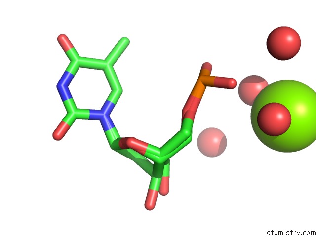
Mono view
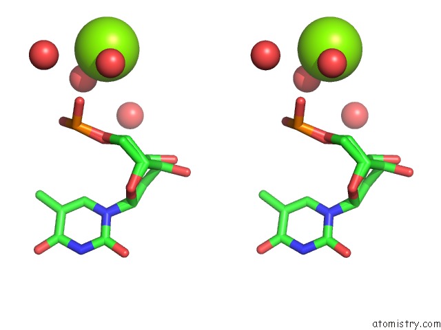
Stereo pair view
Mono view
Stereo pair view
A full contact list of Magnesium with other atoms in the Mg binding
site number 1 of X-Ray Strucutre of An Echinomycin-(Gcgtacgc)2 Complex within 5.0Å range:
|
Reference:
J.A.Cuesta-Seijo,
G.M.Sheldrick.
Structures of Complexes Between Echinomycin and Duplex Dna. Acta Crystallogr.,Sect.D V. 61 442 2005.
ISSN: ISSN 0907-4449
PubMed: 15805599
DOI: 10.1107/S090744490500137X
Page generated: Tue Aug 13 18:24:55 2024
ISSN: ISSN 0907-4449
PubMed: 15805599
DOI: 10.1107/S090744490500137X
Last articles
Zn in 9J0NZn in 9J0O
Zn in 9J0P
Zn in 9FJX
Zn in 9EKB
Zn in 9C0F
Zn in 9CAH
Zn in 9CH0
Zn in 9CH3
Zn in 9CH1