Magnesium »
PDB 1xoq-1xyc »
1xx1 »
Magnesium in PDB 1xx1: Structural Basis For Ion-Coordination and the Catalytic Mechanism of Sphingomyelinases D
Enzymatic activity of Structural Basis For Ion-Coordination and the Catalytic Mechanism of Sphingomyelinases D
All present enzymatic activity of Structural Basis For Ion-Coordination and the Catalytic Mechanism of Sphingomyelinases D:
3.1.4.41;
3.1.4.41;
Protein crystallography data
The structure of Structural Basis For Ion-Coordination and the Catalytic Mechanism of Sphingomyelinases D, PDB code: 1xx1
was solved by
M.T.Murakami,
D.V.Tambourgi,
R.K.Arni,
with X-Ray Crystallography technique. A brief refinement statistics is given in the table below:
| Resolution Low / High (Å) | 30.00 / 1.75 |
| Space group | P 65 |
| Cell size a, b, c (Å), α, β, γ (°) | 139.818, 139.818, 113.461, 90.00, 90.00, 120.00 |
| R / Rfree (%) | 18.6 / 22.5 |
Magnesium Binding Sites:
The binding sites of Magnesium atom in the Structural Basis For Ion-Coordination and the Catalytic Mechanism of Sphingomyelinases D
(pdb code 1xx1). This binding sites where shown within
5.0 Angstroms radius around Magnesium atom.
In total 4 binding sites of Magnesium where determined in the Structural Basis For Ion-Coordination and the Catalytic Mechanism of Sphingomyelinases D, PDB code: 1xx1:
Jump to Magnesium binding site number: 1; 2; 3; 4;
In total 4 binding sites of Magnesium where determined in the Structural Basis For Ion-Coordination and the Catalytic Mechanism of Sphingomyelinases D, PDB code: 1xx1:
Jump to Magnesium binding site number: 1; 2; 3; 4;
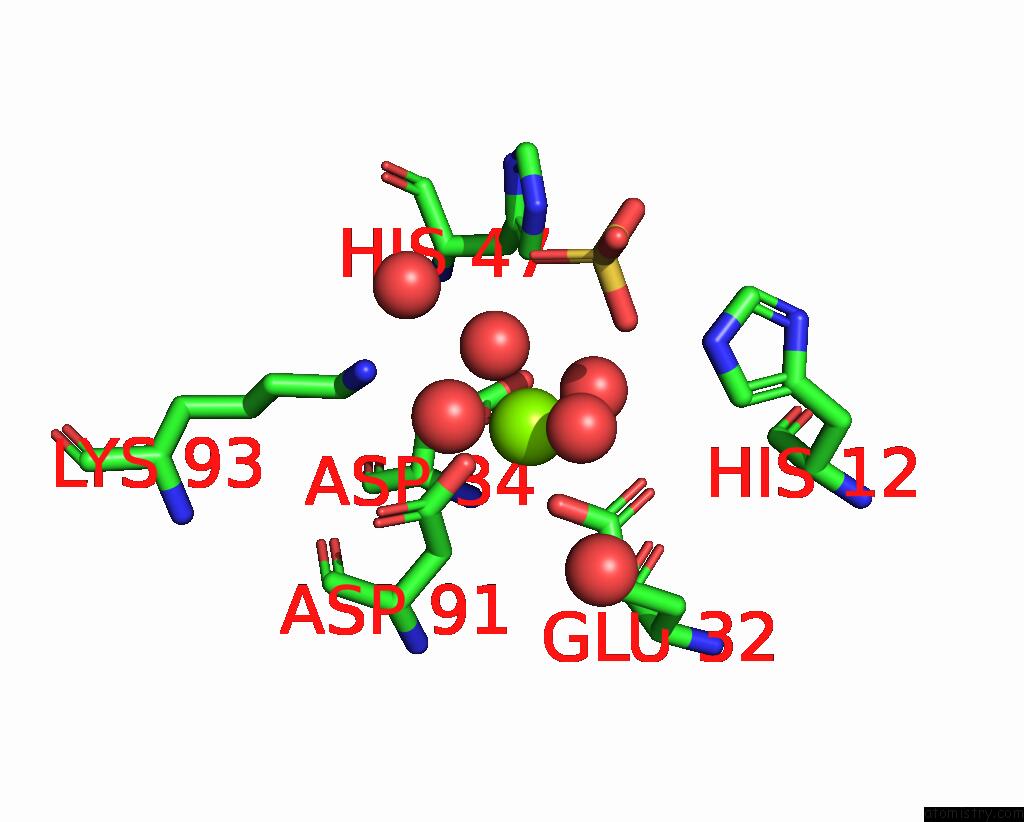
Magnesium binding site 1 out of 4 in 1xx1
Go back to
Magnesium binding site 1 out
of 4 in the Structural Basis For Ion-Coordination and the Catalytic Mechanism of Sphingomyelinases D

Mono view
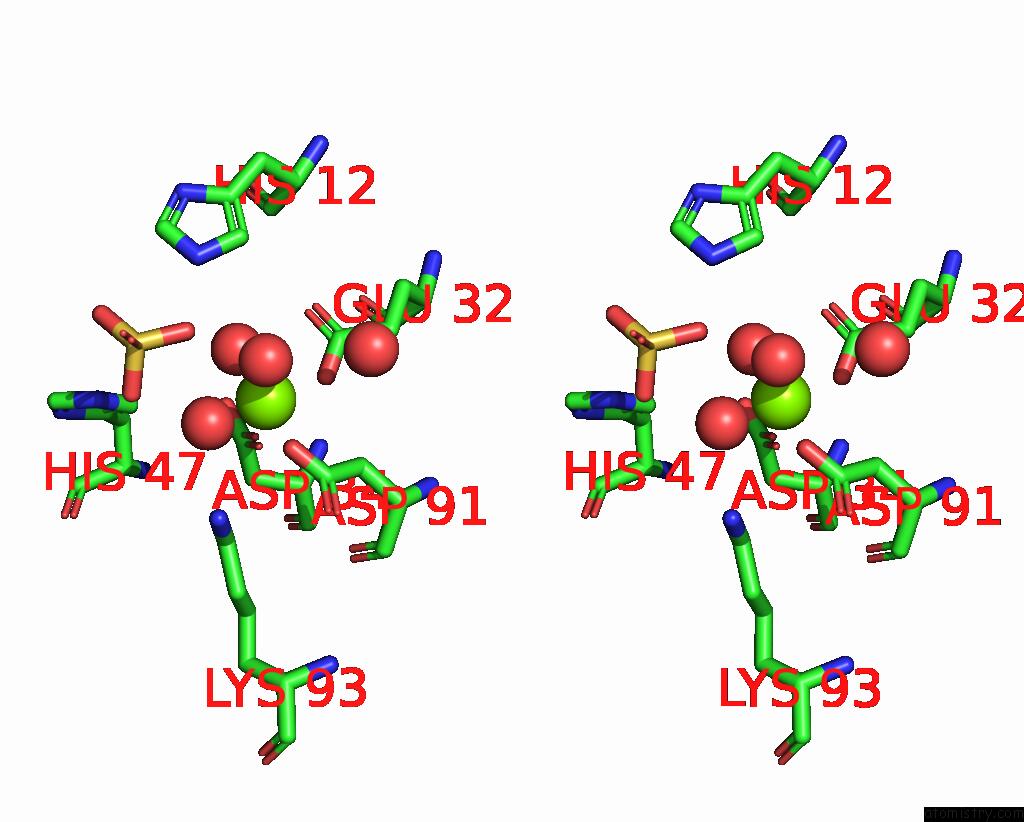
Stereo pair view
Mono view
Stereo pair view
A full contact list of Magnesium with other atoms in the Mg binding
site number 1 of Structural Basis For Ion-Coordination and the Catalytic Mechanism of Sphingomyelinases D within 5.0Å range:
|
Magnesium binding site 2 out of 4 in 1xx1
Go back to
Magnesium binding site 2 out
of 4 in the Structural Basis For Ion-Coordination and the Catalytic Mechanism of Sphingomyelinases D

Mono view

Stereo pair view
Mono view
Stereo pair view
A full contact list of Magnesium with other atoms in the Mg binding
site number 2 of Structural Basis For Ion-Coordination and the Catalytic Mechanism of Sphingomyelinases D within 5.0Å range:
|
Magnesium binding site 3 out of 4 in 1xx1
Go back to
Magnesium binding site 3 out
of 4 in the Structural Basis For Ion-Coordination and the Catalytic Mechanism of Sphingomyelinases D

Mono view

Stereo pair view
Mono view
Stereo pair view
A full contact list of Magnesium with other atoms in the Mg binding
site number 3 of Structural Basis For Ion-Coordination and the Catalytic Mechanism of Sphingomyelinases D within 5.0Å range:
|
Magnesium binding site 4 out of 4 in 1xx1
Go back to
Magnesium binding site 4 out
of 4 in the Structural Basis For Ion-Coordination and the Catalytic Mechanism of Sphingomyelinases D
Mono view

Stereo pair view
Mono view
Stereo pair view
A full contact list of Magnesium with other atoms in the Mg binding
site number 4 of Structural Basis For Ion-Coordination and the Catalytic Mechanism of Sphingomyelinases D within 5.0Å range:
|
Reference:
M.T.Murakami,
M.F.Fernandes-Pedrosa,
D.V.Tambourgi,
R.K.Arni.
Structural Basis For Metal Ion Coordination and the Catalytic Mechanism of Sphingomyelinases D J.Biol.Chem. V. 280 13658 2005.
ISSN: ISSN 0021-9258
PubMed: 15654080
DOI: 10.1074/JBC.M412437200
Page generated: Tue Aug 13 18:25:19 2024
ISSN: ISSN 0021-9258
PubMed: 15654080
DOI: 10.1074/JBC.M412437200
Last articles
Fe in 2YXOFe in 2YRS
Fe in 2YXC
Fe in 2YNM
Fe in 2YVJ
Fe in 2YP1
Fe in 2YU2
Fe in 2YU1
Fe in 2YQB
Fe in 2YOO