Magnesium »
PDB 1ye8-1yqw »
1yjn »
Magnesium in PDB 1yjn: Crystal Structure of Clindamycin Bound to the G2099A Mutant 50S Ribosomal Subunit of Haloarcula Marismortui
Protein crystallography data
The structure of Crystal Structure of Clindamycin Bound to the G2099A Mutant 50S Ribosomal Subunit of Haloarcula Marismortui, PDB code: 1yjn
was solved by
D.Tu,
G.Blaha,
P.B.Moore,
T.A.Steitz,
with X-Ray Crystallography technique. A brief refinement statistics is given in the table below:
| Resolution Low / High (Å) | 29.97 / 3.00 |
| Space group | C 2 2 21 |
| Cell size a, b, c (Å), α, β, γ (°) | 212.512, 300.125, 573.849, 90.00, 90.00, 90.00 |
| R / Rfree (%) | 17 / 22.8 |
Other elements in 1yjn:
The structure of Crystal Structure of Clindamycin Bound to the G2099A Mutant 50S Ribosomal Subunit of Haloarcula Marismortui also contains other interesting chemical elements:
| Potassium | (K) | 2 atoms |
| Cadmium | (Cd) | 5 atoms |
| Chlorine | (Cl) | 23 atoms |
| Sodium | (Na) | 86 atoms |
Magnesium Binding Sites:
Pages:
>>> Page 1 <<< Page 2, Binding sites: 11 - 20; Page 3, Binding sites: 21 - 30; Page 4, Binding sites: 31 - 40; Page 5, Binding sites: 41 - 50; Page 6, Binding sites: 51 - 60; Page 7, Binding sites: 61 - 70; Page 8, Binding sites: 71 - 80; Page 9, Binding sites: 81 - 90; Page 10, Binding sites: 91 - 100; Page 11, Binding sites: 101 - 110; Page 12, Binding sites: 111 - 116;Binding sites:
The binding sites of Magnesium atom in the Crystal Structure of Clindamycin Bound to the G2099A Mutant 50S Ribosomal Subunit of Haloarcula Marismortui (pdb code 1yjn). This binding sites where shown within 5.0 Angstroms radius around Magnesium atom.In total 116 binding sites of Magnesium where determined in the Crystal Structure of Clindamycin Bound to the G2099A Mutant 50S Ribosomal Subunit of Haloarcula Marismortui, PDB code: 1yjn:
Jump to Magnesium binding site number: 1; 2; 3; 4; 5; 6; 7; 8; 9; 10;
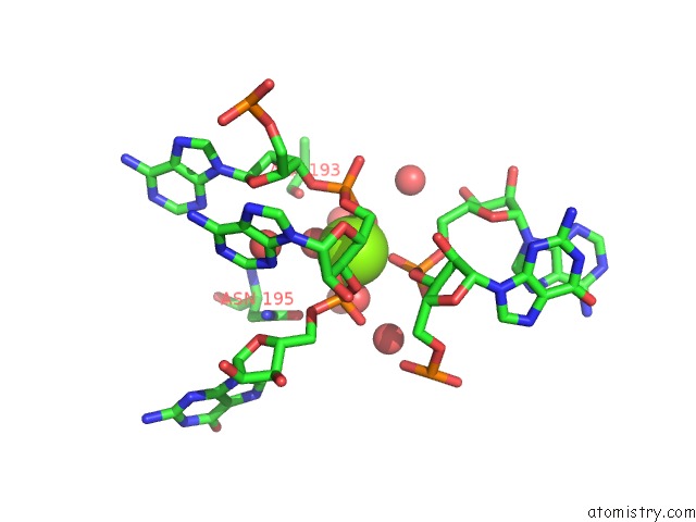
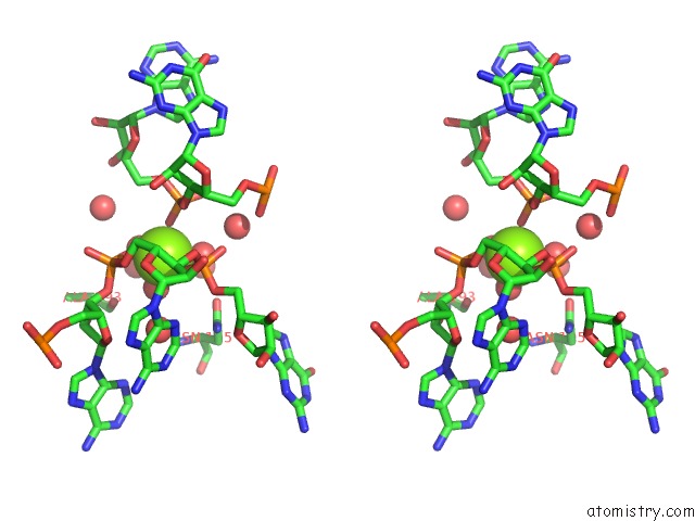
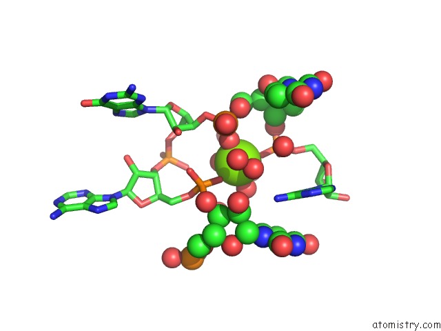
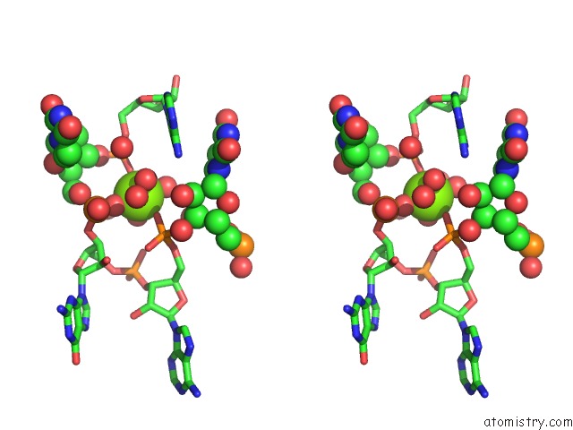
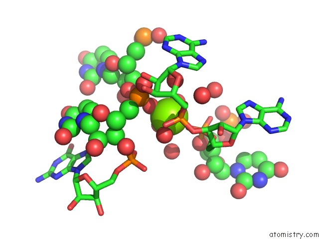
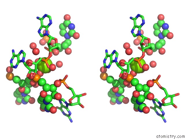
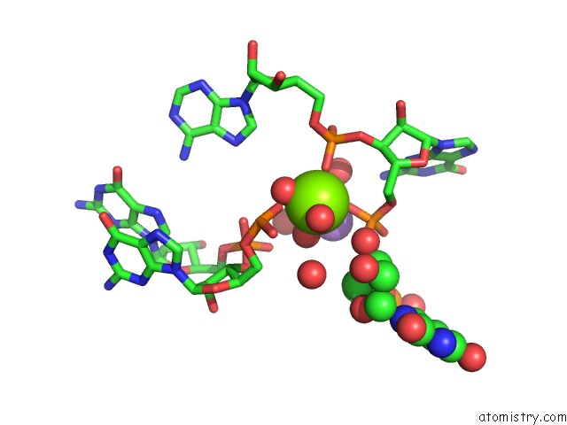
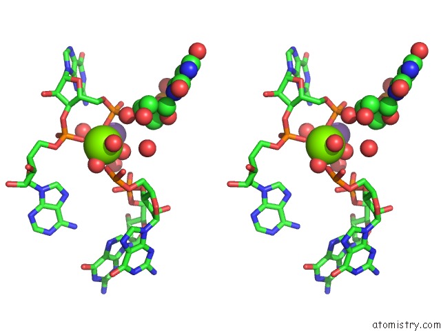
Magnesium binding site 1 out of 116 in 1yjn
Go back to
Magnesium binding site 1 out
of 116 in the Crystal Structure of Clindamycin Bound to the G2099A Mutant 50S Ribosomal Subunit of Haloarcula Marismortui
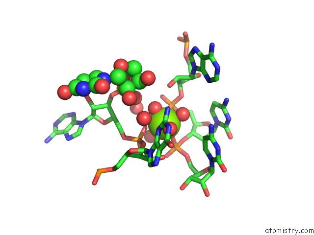
Mono view
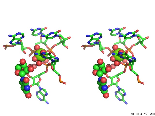
Stereo pair view
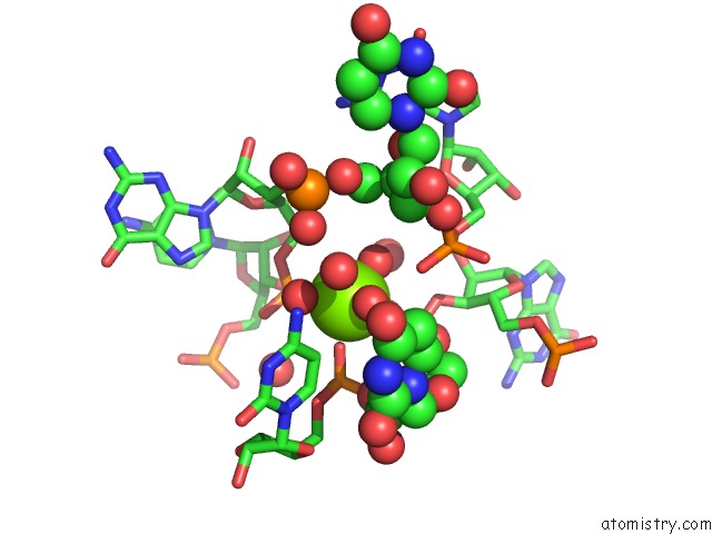
Mono view
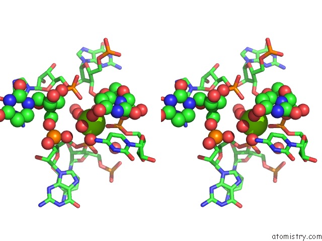
Stereo pair view
A full contact list of Magnesium with other atoms in the Mg binding
site number 1 of Crystal Structure of Clindamycin Bound to the G2099A Mutant 50S Ribosomal Subunit of Haloarcula Marismortui within 5.0Å range:
|
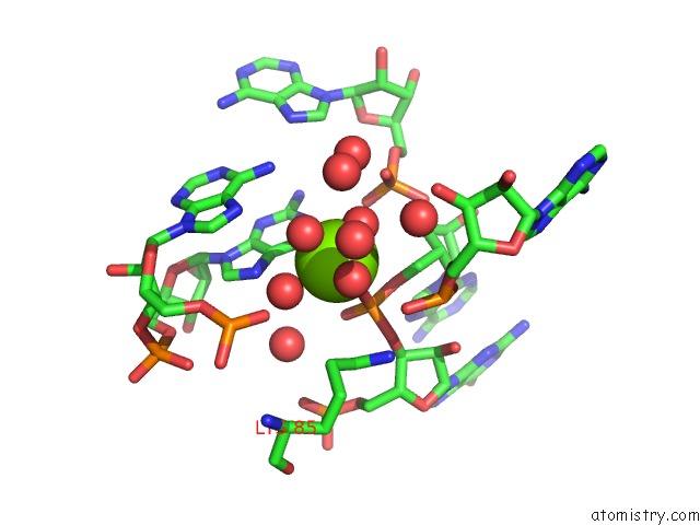
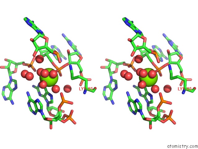
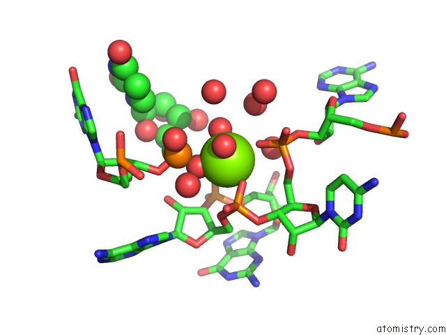
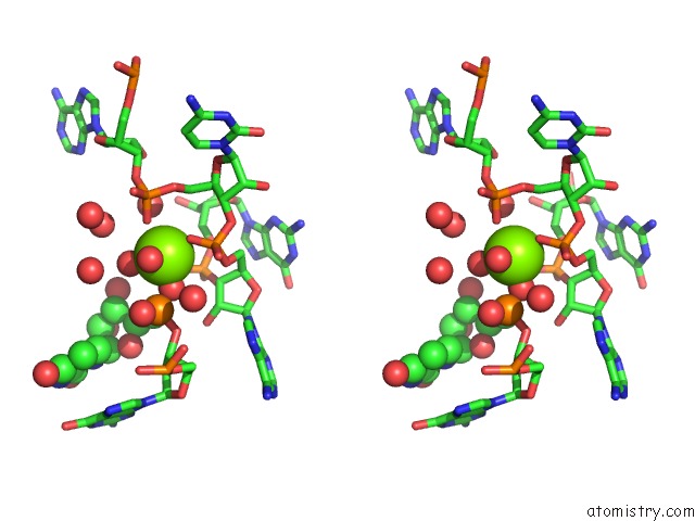
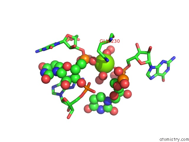
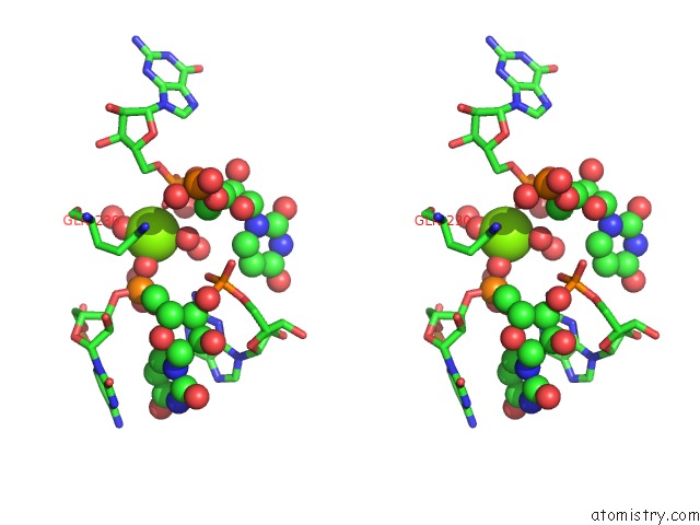
Magnesium binding site 2 out of 116 in 1yjn
Go back to
Magnesium binding site 2 out
of 116 in the Crystal Structure of Clindamycin Bound to the G2099A Mutant 50S Ribosomal Subunit of Haloarcula Marismortui
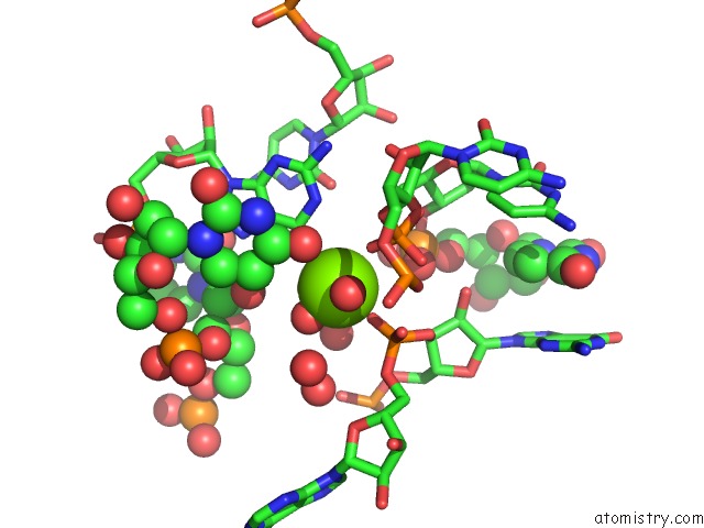
Mono view
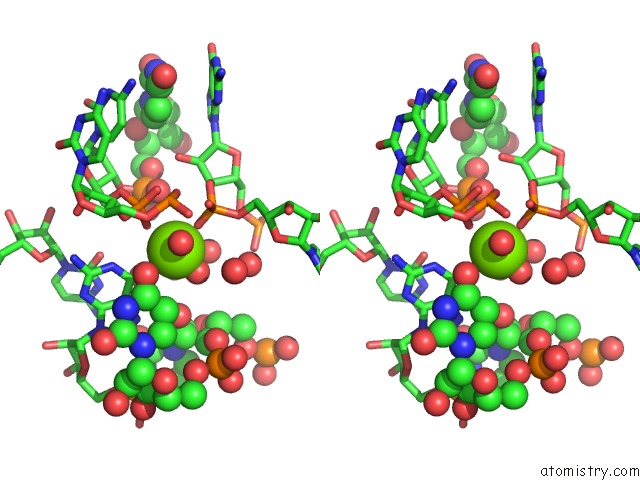
Stereo pair view
Mono view
Stereo pair view
A full contact list of Magnesium with other atoms in the Mg binding
site number 2 of Crystal Structure of Clindamycin Bound to the G2099A Mutant 50S Ribosomal Subunit of Haloarcula Marismortui within 5.0Å range:
|
Magnesium binding site 3 out of 116 in 1yjn
Go back to
Magnesium binding site 3 out
of 116 in the Crystal Structure of Clindamycin Bound to the G2099A Mutant 50S Ribosomal Subunit of Haloarcula Marismortui
Mono view
Stereo pair view
Mono view
Stereo pair view
A full contact list of Magnesium with other atoms in the Mg binding
site number 3 of Crystal Structure of Clindamycin Bound to the G2099A Mutant 50S Ribosomal Subunit of Haloarcula Marismortui within 5.0Å range:
|
Magnesium binding site 4 out of 116 in 1yjn
Go back to
Magnesium binding site 4 out
of 116 in the Crystal Structure of Clindamycin Bound to the G2099A Mutant 50S Ribosomal Subunit of Haloarcula Marismortui
Mono view
Stereo pair view
Mono view
Stereo pair view
A full contact list of Magnesium with other atoms in the Mg binding
site number 4 of Crystal Structure of Clindamycin Bound to the G2099A Mutant 50S Ribosomal Subunit of Haloarcula Marismortui within 5.0Å range:
|
Magnesium binding site 5 out of 116 in 1yjn
Go back to
Magnesium binding site 5 out
of 116 in the Crystal Structure of Clindamycin Bound to the G2099A Mutant 50S Ribosomal Subunit of Haloarcula Marismortui
Mono view
Stereo pair view
Mono view
Stereo pair view
A full contact list of Magnesium with other atoms in the Mg binding
site number 5 of Crystal Structure of Clindamycin Bound to the G2099A Mutant 50S Ribosomal Subunit of Haloarcula Marismortui within 5.0Å range:
|
Magnesium binding site 6 out of 116 in 1yjn
Go back to
Magnesium binding site 6 out
of 116 in the Crystal Structure of Clindamycin Bound to the G2099A Mutant 50S Ribosomal Subunit of Haloarcula Marismortui
Mono view
Stereo pair view
Mono view
Stereo pair view
A full contact list of Magnesium with other atoms in the Mg binding
site number 6 of Crystal Structure of Clindamycin Bound to the G2099A Mutant 50S Ribosomal Subunit of Haloarcula Marismortui within 5.0Å range:
|
Magnesium binding site 7 out of 116 in 1yjn
Go back to
Magnesium binding site 7 out
of 116 in the Crystal Structure of Clindamycin Bound to the G2099A Mutant 50S Ribosomal Subunit of Haloarcula Marismortui
Mono view
Stereo pair view
Mono view
Stereo pair view
A full contact list of Magnesium with other atoms in the Mg binding
site number 7 of Crystal Structure of Clindamycin Bound to the G2099A Mutant 50S Ribosomal Subunit of Haloarcula Marismortui within 5.0Å range:
|
Magnesium binding site 8 out of 116 in 1yjn
Go back to
Magnesium binding site 8 out
of 116 in the Crystal Structure of Clindamycin Bound to the G2099A Mutant 50S Ribosomal Subunit of Haloarcula Marismortui
Mono view
Stereo pair view
Mono view
Stereo pair view
A full contact list of Magnesium with other atoms in the Mg binding
site number 8 of Crystal Structure of Clindamycin Bound to the G2099A Mutant 50S Ribosomal Subunit of Haloarcula Marismortui within 5.0Å range:
|
Magnesium binding site 9 out of 116 in 1yjn
Go back to
Magnesium binding site 9 out
of 116 in the Crystal Structure of Clindamycin Bound to the G2099A Mutant 50S Ribosomal Subunit of Haloarcula Marismortui
Mono view
Stereo pair view
Mono view
Stereo pair view
A full contact list of Magnesium with other atoms in the Mg binding
site number 9 of Crystal Structure of Clindamycin Bound to the G2099A Mutant 50S Ribosomal Subunit of Haloarcula Marismortui within 5.0Å range:
|
Magnesium binding site 10 out of 116 in 1yjn
Go back to
Magnesium binding site 10 out
of 116 in the Crystal Structure of Clindamycin Bound to the G2099A Mutant 50S Ribosomal Subunit of Haloarcula Marismortui
Mono view
Stereo pair view
Mono view
Stereo pair view
A full contact list of Magnesium with other atoms in the Mg binding
site number 10 of Crystal Structure of Clindamycin Bound to the G2099A Mutant 50S Ribosomal Subunit of Haloarcula Marismortui within 5.0Å range:
|
Reference:
D.Tu,
G.Blaha,
P.B.Moore,
T.A.Steitz.
Structures of Mlsbk Antibiotics Bound to Mutated Large Ribosomal Subunits Provide A Structural Explanation For Resistance. Cell(Cambridge,Mass.) V. 121 257 2005.
ISSN: ISSN 0092-8674
PubMed: 15851032
DOI: 10.1016/J.CELL.2005.02.005
Page generated: Tue Aug 13 18:42:38 2024
ISSN: ISSN 0092-8674
PubMed: 15851032
DOI: 10.1016/J.CELL.2005.02.005
Last articles
Fe in 2YXOFe in 2YRS
Fe in 2YXC
Fe in 2YNM
Fe in 2YVJ
Fe in 2YP1
Fe in 2YU2
Fe in 2YU1
Fe in 2YQB
Fe in 2YOO