Magnesium »
PDB 1ye8-1yqw »
1ykq »
Magnesium in PDB 1ykq: Crystal Structure of Diels-Alder Ribozyme
Protein crystallography data
The structure of Crystal Structure of Diels-Alder Ribozyme, PDB code: 1ykq
was solved by
A.Serganov,
S.Keiper,
L.Malinina,
V.Tereshko,
E.Skripkin,
C.Hobartner,
A.Polonskaia,
A.T.Phan,
R.Wombacher,
R.Micura,
Z.Dauter,
A.Jaschke,
D.J.Patel,
with X-Ray Crystallography technique. A brief refinement statistics is given in the table below:
| Resolution Low / High (Å) | 20.00 / 3.50 |
| Space group | P 1 21 1 |
| Cell size a, b, c (Å), α, β, γ (°) | 77.690, 44.240, 79.640, 90.00, 107.12, 90.00 |
| R / Rfree (%) | 28.6 / 32.4 |
Other elements in 1ykq:
The structure of Crystal Structure of Diels-Alder Ribozyme also contains other interesting chemical elements:
| Cadmium | (Cd) | 5 atoms |
Magnesium Binding Sites:
The binding sites of Magnesium atom in the Crystal Structure of Diels-Alder Ribozyme
(pdb code 1ykq). This binding sites where shown within
5.0 Angstroms radius around Magnesium atom.
In total 7 binding sites of Magnesium where determined in the Crystal Structure of Diels-Alder Ribozyme, PDB code: 1ykq:
Jump to Magnesium binding site number: 1; 2; 3; 4; 5; 6; 7;
In total 7 binding sites of Magnesium where determined in the Crystal Structure of Diels-Alder Ribozyme, PDB code: 1ykq:
Jump to Magnesium binding site number: 1; 2; 3; 4; 5; 6; 7;
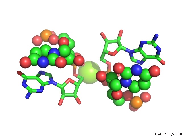
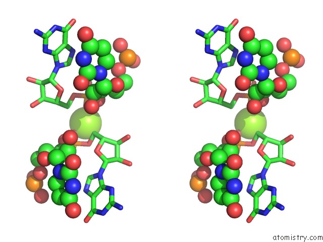
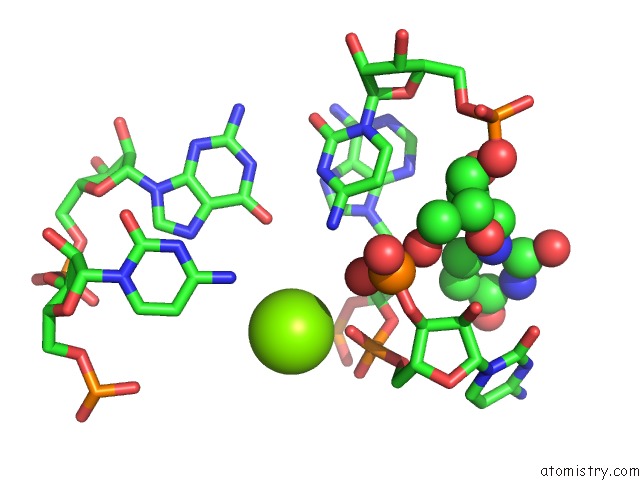
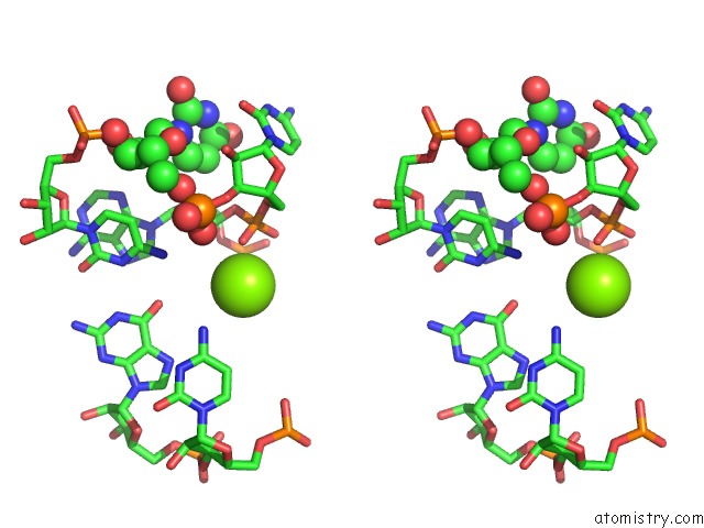
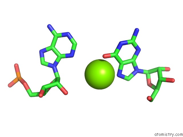
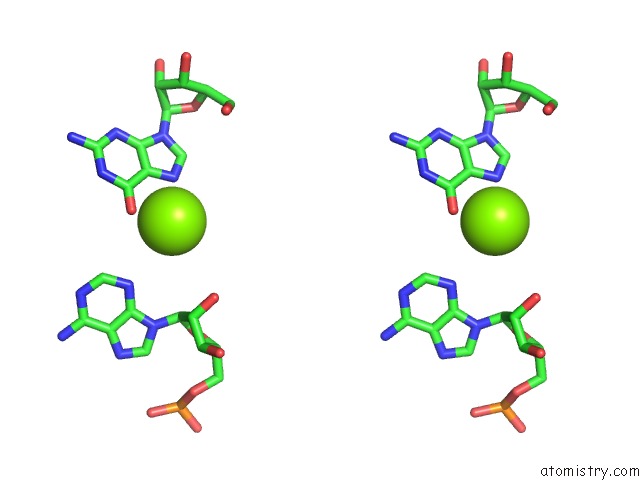
Magnesium binding site 1 out of 7 in 1ykq
Go back to
Magnesium binding site 1 out
of 7 in the Crystal Structure of Diels-Alder Ribozyme
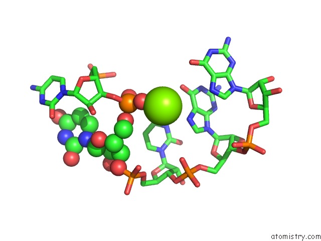
Mono view
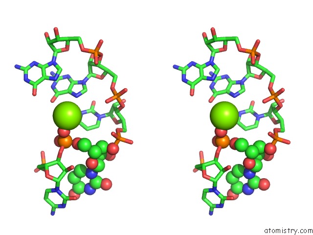
Stereo pair view
Mono view
Stereo pair view
A full contact list of Magnesium with other atoms in the Mg binding
site number 1 of Crystal Structure of Diels-Alder Ribozyme within 5.0Å range:
|
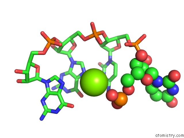
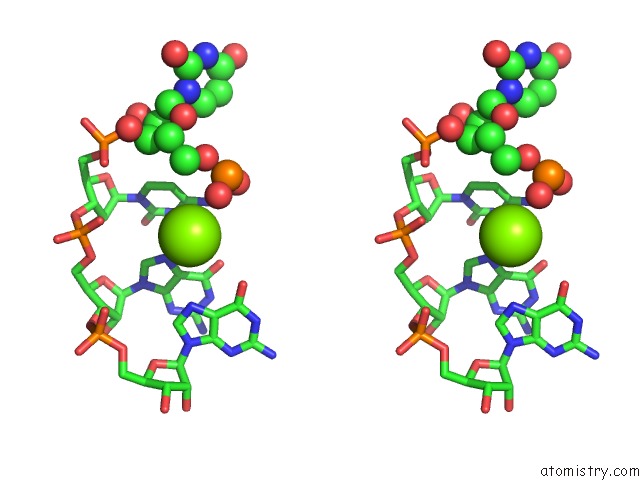
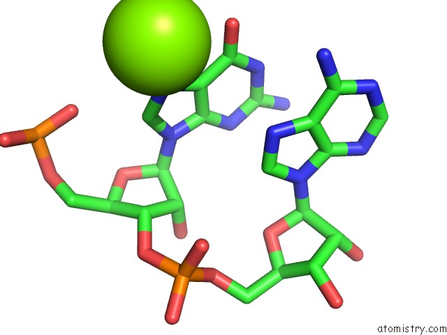
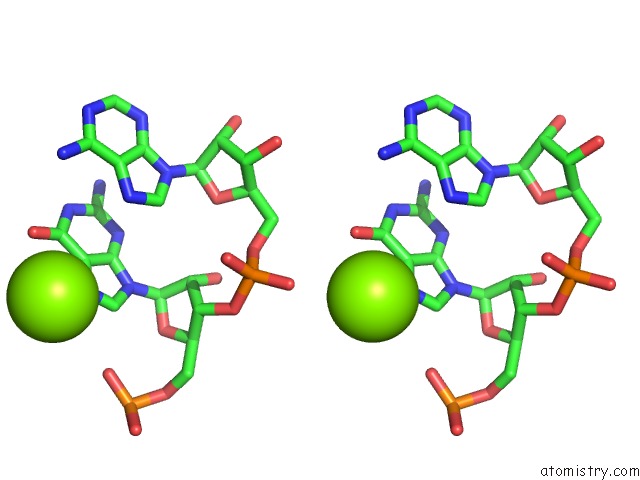
Magnesium binding site 2 out of 7 in 1ykq
Go back to
Magnesium binding site 2 out
of 7 in the Crystal Structure of Diels-Alder Ribozyme
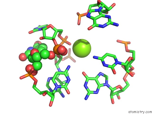
Mono view
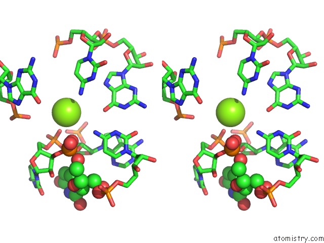
Stereo pair view
Mono view
Stereo pair view
A full contact list of Magnesium with other atoms in the Mg binding
site number 2 of Crystal Structure of Diels-Alder Ribozyme within 5.0Å range:
|
Magnesium binding site 3 out of 7 in 1ykq
Go back to
Magnesium binding site 3 out
of 7 in the Crystal Structure of Diels-Alder Ribozyme
Mono view
Stereo pair view
Mono view
Stereo pair view
A full contact list of Magnesium with other atoms in the Mg binding
site number 3 of Crystal Structure of Diels-Alder Ribozyme within 5.0Å range:
|
Magnesium binding site 4 out of 7 in 1ykq
Go back to
Magnesium binding site 4 out
of 7 in the Crystal Structure of Diels-Alder Ribozyme
Mono view
Stereo pair view
Mono view
Stereo pair view
A full contact list of Magnesium with other atoms in the Mg binding
site number 4 of Crystal Structure of Diels-Alder Ribozyme within 5.0Å range:
|
Magnesium binding site 5 out of 7 in 1ykq
Go back to
Magnesium binding site 5 out
of 7 in the Crystal Structure of Diels-Alder Ribozyme
Mono view
Stereo pair view
Mono view
Stereo pair view
A full contact list of Magnesium with other atoms in the Mg binding
site number 5 of Crystal Structure of Diels-Alder Ribozyme within 5.0Å range:
|
Magnesium binding site 6 out of 7 in 1ykq
Go back to
Magnesium binding site 6 out
of 7 in the Crystal Structure of Diels-Alder Ribozyme
Mono view
Stereo pair view
Mono view
Stereo pair view
A full contact list of Magnesium with other atoms in the Mg binding
site number 6 of Crystal Structure of Diels-Alder Ribozyme within 5.0Å range:
|
Magnesium binding site 7 out of 7 in 1ykq
Go back to
Magnesium binding site 7 out
of 7 in the Crystal Structure of Diels-Alder Ribozyme
Mono view
Stereo pair view
Mono view
Stereo pair view
A full contact list of Magnesium with other atoms in the Mg binding
site number 7 of Crystal Structure of Diels-Alder Ribozyme within 5.0Å range:
|
Reference:
A.Serganov,
S.Keiper,
L.Malinina,
V.Tereshko,
E.Skripkin,
C.Hobartner,
A.Polonskaia,
A.T.Phan,
R.Wombacher,
R.Micura,
Z.Dauter,
A.Jaschke,
D.J.Patel.
Structural Basis For Diels-Alder Ribozyme-Catalyzed Carbon-Carbon Bond Formation. Nat.Struct.Mol.Biol. V. 12 218 2005.
ISSN: ISSN 1545-9993
PubMed: 15723077
DOI: 10.1038/NSMB906
Page generated: Tue Aug 13 19:35:58 2024
ISSN: ISSN 1545-9993
PubMed: 15723077
DOI: 10.1038/NSMB906
Last articles
Zn in 9MJ5Zn in 9HNW
Zn in 9G0L
Zn in 9FNE
Zn in 9DZN
Zn in 9E0I
Zn in 9D32
Zn in 9DAK
Zn in 8ZXC
Zn in 8ZUF