Magnesium »
PDB 1ye8-1yqw »
1yl5 »
Magnesium in PDB 1yl5: Crystal Structure of Mycobacterium Tuberculosis Dihydrodipicolinate Reductase (RV2773C) (Crystal Form A)
Enzymatic activity of Crystal Structure of Mycobacterium Tuberculosis Dihydrodipicolinate Reductase (RV2773C) (Crystal Form A)
All present enzymatic activity of Crystal Structure of Mycobacterium Tuberculosis Dihydrodipicolinate Reductase (RV2773C) (Crystal Form A):
1.3.1.26;
1.3.1.26;
Protein crystallography data
The structure of Crystal Structure of Mycobacterium Tuberculosis Dihydrodipicolinate Reductase (RV2773C) (Crystal Form A), PDB code: 1yl5
was solved by
R.Janowski,
G.Kefala,
M.S.Weiss,
Tb Structural Genomics Consortium(Tbsgc),
with X-Ray Crystallography technique. A brief refinement statistics is given in the table below:
| Resolution Low / High (Å) | 30.00 / 2.30 |
| Space group | P 21 21 2 |
| Cell size a, b, c (Å), α, β, γ (°) | 59.260, 122.000, 77.930, 90.00, 90.00, 90.00 |
| R / Rfree (%) | 20.5 / 24.9 |
Magnesium Binding Sites:
The binding sites of Magnesium atom in the Crystal Structure of Mycobacterium Tuberculosis Dihydrodipicolinate Reductase (RV2773C) (Crystal Form A)
(pdb code 1yl5). This binding sites where shown within
5.0 Angstroms radius around Magnesium atom.
In total 2 binding sites of Magnesium where determined in the Crystal Structure of Mycobacterium Tuberculosis Dihydrodipicolinate Reductase (RV2773C) (Crystal Form A), PDB code: 1yl5:
Jump to Magnesium binding site number: 1; 2;
In total 2 binding sites of Magnesium where determined in the Crystal Structure of Mycobacterium Tuberculosis Dihydrodipicolinate Reductase (RV2773C) (Crystal Form A), PDB code: 1yl5:
Jump to Magnesium binding site number: 1; 2;
Magnesium binding site 1 out of 2 in 1yl5
Go back to
Magnesium binding site 1 out
of 2 in the Crystal Structure of Mycobacterium Tuberculosis Dihydrodipicolinate Reductase (RV2773C) (Crystal Form A)
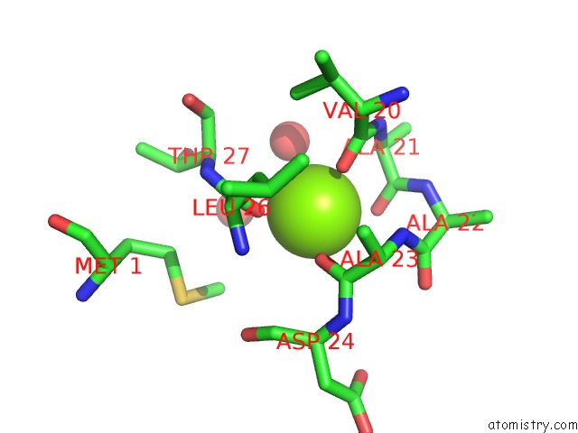
Mono view
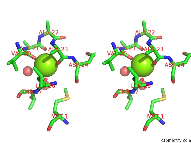
Stereo pair view
Mono view
Stereo pair view
A full contact list of Magnesium with other atoms in the Mg binding
site number 1 of Crystal Structure of Mycobacterium Tuberculosis Dihydrodipicolinate Reductase (RV2773C) (Crystal Form A) within 5.0Å range:
|
Magnesium binding site 2 out of 2 in 1yl5
Go back to
Magnesium binding site 2 out
of 2 in the Crystal Structure of Mycobacterium Tuberculosis Dihydrodipicolinate Reductase (RV2773C) (Crystal Form A)
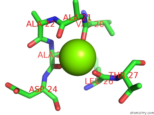
Mono view
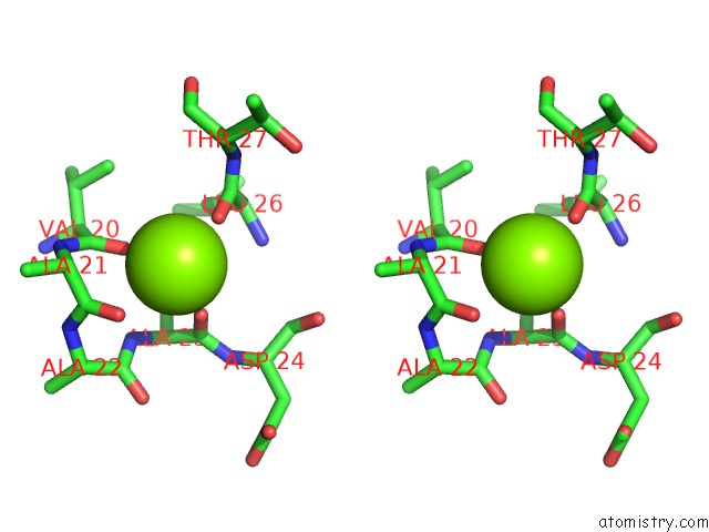
Stereo pair view
Mono view
Stereo pair view
A full contact list of Magnesium with other atoms in the Mg binding
site number 2 of Crystal Structure of Mycobacterium Tuberculosis Dihydrodipicolinate Reductase (RV2773C) (Crystal Form A) within 5.0Å range:
|
Reference:
R.Janowski,
G.Kefala,
M.S.Weiss.
The Structure of Dihydrodipicolinate Reductase (Dapb) From Mycobacterium Tuberculosis in Three Crystal Forms. Acta Crystallogr.,Sect.D V. 66 61 2010.
ISSN: ISSN 0907-4449
PubMed: 20057050
DOI: 10.1107/S0907444909043960
Page generated: Tue Aug 13 19:36:55 2024
ISSN: ISSN 0907-4449
PubMed: 20057050
DOI: 10.1107/S0907444909043960
Last articles
Zn in 9J0NZn in 9J0O
Zn in 9J0P
Zn in 9FJX
Zn in 9EKB
Zn in 9C0F
Zn in 9CAH
Zn in 9CH0
Zn in 9CH3
Zn in 9CH1