Magnesium »
PDB 1yqz-1yzt »
1yrj »
Magnesium in PDB 1yrj: Crystal Structure of Apramycin Bound to A Ribosomal Rna A Site Oligonucleotide
Protein crystallography data
The structure of Crystal Structure of Apramycin Bound to A Ribosomal Rna A Site Oligonucleotide, PDB code: 1yrj
was solved by
Q.Han,
Q.Zhao,
S.Fish,
K.B.Simonsen,
D.Vourloumis,
J.M.Froelich,
D.Wall,
T.Hermann,
with X-Ray Crystallography technique. A brief refinement statistics is given in the table below:
| Resolution Low / High (Å) | 8.00 / 2.70 |
| Space group | P 21 21 2 |
| Cell size a, b, c (Å), α, β, γ (°) | 92.250, 30.900, 45.810, 90.00, 90.00, 90.00 |
| R / Rfree (%) | 24.6 / 30.7 |
Other elements in 1yrj:
The structure of Crystal Structure of Apramycin Bound to A Ribosomal Rna A Site Oligonucleotide also contains other interesting chemical elements:
| Potassium | (K) | 1 atom |
Magnesium Binding Sites:
The binding sites of Magnesium atom in the Crystal Structure of Apramycin Bound to A Ribosomal Rna A Site Oligonucleotide
(pdb code 1yrj). This binding sites where shown within
5.0 Angstroms radius around Magnesium atom.
In total only one binding site of Magnesium was determined in the Crystal Structure of Apramycin Bound to A Ribosomal Rna A Site Oligonucleotide, PDB code: 1yrj:
In total only one binding site of Magnesium was determined in the Crystal Structure of Apramycin Bound to A Ribosomal Rna A Site Oligonucleotide, PDB code: 1yrj:
Magnesium binding site 1 out of 1 in 1yrj
Go back to
Magnesium binding site 1 out
of 1 in the Crystal Structure of Apramycin Bound to A Ribosomal Rna A Site Oligonucleotide
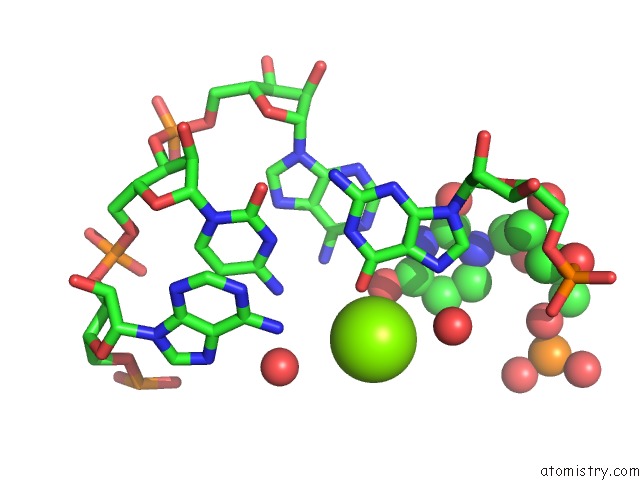
Mono view
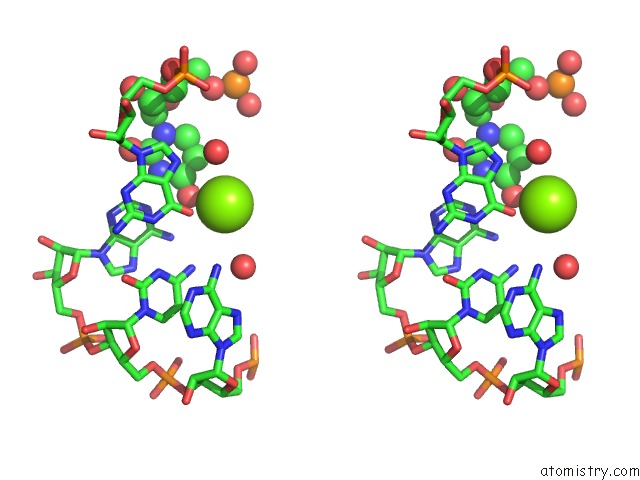
Stereo pair view
Mono view
Stereo pair view
A full contact list of Magnesium with other atoms in the Mg binding
site number 1 of Crystal Structure of Apramycin Bound to A Ribosomal Rna A Site Oligonucleotide within 5.0Å range:
|
Reference:
Q.Han,
Q.Zhao,
S.Fish,
K.B.Simonsen,
D.Vourloumis,
J.M.Froelich,
D.Wall,
T.Hermann.
Molecular Recognition By Glycoside Pseudo Base Pairs and Triples in An Apramycin-Rna Complex. Angew.Chem.Int.Ed.Engl. V. 44 2694 2005.
ISSN: ISSN 1433-7851
PubMed: 15849690
DOI: 10.1002/ANIE.200500028
Page generated: Tue Aug 13 19:52:19 2024
ISSN: ISSN 1433-7851
PubMed: 15849690
DOI: 10.1002/ANIE.200500028
Last articles
Zn in 9J0NZn in 9J0O
Zn in 9J0P
Zn in 9FJX
Zn in 9EKB
Zn in 9C0F
Zn in 9CAH
Zn in 9CH0
Zn in 9CH3
Zn in 9CH1