Magnesium »
PDB 1yqz-1yzt »
1yun »
Magnesium in PDB 1yun: Crystal Structure of Nicotinic Acid Mononucleotide Adenylyltransferase From Pseudomonas Aeruginosa
Enzymatic activity of Crystal Structure of Nicotinic Acid Mononucleotide Adenylyltransferase From Pseudomonas Aeruginosa
All present enzymatic activity of Crystal Structure of Nicotinic Acid Mononucleotide Adenylyltransferase From Pseudomonas Aeruginosa:
2.7.7.18;
2.7.7.18;
Protein crystallography data
The structure of Crystal Structure of Nicotinic Acid Mononucleotide Adenylyltransferase From Pseudomonas Aeruginosa, PDB code: 1yun
was solved by
H.J.Yoon,
H.L.Kim,
B.Mikami,
S.W.Suh,
with X-Ray Crystallography technique. A brief refinement statistics is given in the table below:
| Resolution Low / High (Å) | 15.00 / 2.00 |
| Space group | P 2 2 21 |
| Cell size a, b, c (Å), α, β, γ (°) | 65.189, 65.199, 110.121, 90.00, 90.00, 90.00 |
| R / Rfree (%) | n/a / 26.9 |
Magnesium Binding Sites:
The binding sites of Magnesium atom in the Crystal Structure of Nicotinic Acid Mononucleotide Adenylyltransferase From Pseudomonas Aeruginosa
(pdb code 1yun). This binding sites where shown within
5.0 Angstroms radius around Magnesium atom.
In total only one binding site of Magnesium was determined in the Crystal Structure of Nicotinic Acid Mononucleotide Adenylyltransferase From Pseudomonas Aeruginosa, PDB code: 1yun:
In total only one binding site of Magnesium was determined in the Crystal Structure of Nicotinic Acid Mononucleotide Adenylyltransferase From Pseudomonas Aeruginosa, PDB code: 1yun:
Magnesium binding site 1 out of 1 in 1yun
Go back to
Magnesium binding site 1 out
of 1 in the Crystal Structure of Nicotinic Acid Mononucleotide Adenylyltransferase From Pseudomonas Aeruginosa
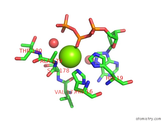
Mono view
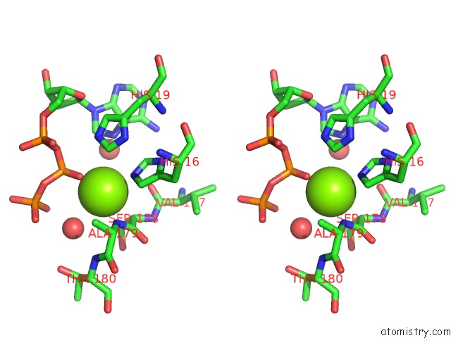
Stereo pair view
Mono view
Stereo pair view
A full contact list of Magnesium with other atoms in the Mg binding
site number 1 of Crystal Structure of Nicotinic Acid Mononucleotide Adenylyltransferase From Pseudomonas Aeruginosa within 5.0Å range:
|
Reference:
H.J.Yoon,
H.L.Kim,
B.Mikami,
S.W.Suh.
Crystal Structure of Nicotinic Acid Mononucleotide Adenylyltransferase From Pseudomonas Aeruginosa in Its Apo and Substrate-Complexed Forms Reveals A Fully Open Conformation J.Mol.Biol. V. 351 258 2005.
ISSN: ISSN 0022-2836
PubMed: 16009375
DOI: 10.1016/J.JMB.2005.06.001
Page generated: Tue Aug 13 19:54:02 2024
ISSN: ISSN 0022-2836
PubMed: 16009375
DOI: 10.1016/J.JMB.2005.06.001
Last articles
Zn in 9J0NZn in 9J0O
Zn in 9J0P
Zn in 9FJX
Zn in 9EKB
Zn in 9C0F
Zn in 9CAH
Zn in 9CH0
Zn in 9CH3
Zn in 9CH1