Magnesium »
PDB 1yqz-1yzt »
1yw0 »
Magnesium in PDB 1yw0: Crystal Structure of the Tryptophan 2,3-Dioxygenase From Xanthomonas Campestris. Northeast Structural Genomics Target XCR13.
Enzymatic activity of Crystal Structure of the Tryptophan 2,3-Dioxygenase From Xanthomonas Campestris. Northeast Structural Genomics Target XCR13.
All present enzymatic activity of Crystal Structure of the Tryptophan 2,3-Dioxygenase From Xanthomonas Campestris. Northeast Structural Genomics Target XCR13.:
1.13.11.11;
1.13.11.11;
Protein crystallography data
The structure of Crystal Structure of the Tryptophan 2,3-Dioxygenase From Xanthomonas Campestris. Northeast Structural Genomics Target XCR13., PDB code: 1yw0
was solved by
S.M.Vorobiev,
M.Abashidze,
F.Forouhar,
A.Kuzin,
R.Xiao,
M.Ciano,
T.B.Aton,
G.T.Montelione,
J.F.Hunt,
L.Tong,
Northeaststructural Genomics Consortium (Nesg),
with X-Ray Crystallography technique. A brief refinement statistics is given in the table below:
| Resolution Low / High (Å) | 29.32 / 2.70 |
| Space group | P 21 21 21 |
| Cell size a, b, c (Å), α, β, γ (°) | 89.701, 110.223, 149.398, 90.00, 90.00, 90.00 |
| R / Rfree (%) | 25 / 29.4 |
Magnesium Binding Sites:
The binding sites of Magnesium atom in the Crystal Structure of the Tryptophan 2,3-Dioxygenase From Xanthomonas Campestris. Northeast Structural Genomics Target XCR13.
(pdb code 1yw0). This binding sites where shown within
5.0 Angstroms radius around Magnesium atom.
In total 8 binding sites of Magnesium where determined in the Crystal Structure of the Tryptophan 2,3-Dioxygenase From Xanthomonas Campestris. Northeast Structural Genomics Target XCR13., PDB code: 1yw0:
Jump to Magnesium binding site number: 1; 2; 3; 4; 5; 6; 7; 8;
In total 8 binding sites of Magnesium where determined in the Crystal Structure of the Tryptophan 2,3-Dioxygenase From Xanthomonas Campestris. Northeast Structural Genomics Target XCR13., PDB code: 1yw0:
Jump to Magnesium binding site number: 1; 2; 3; 4; 5; 6; 7; 8;
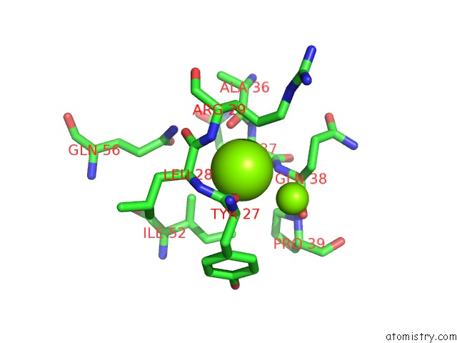
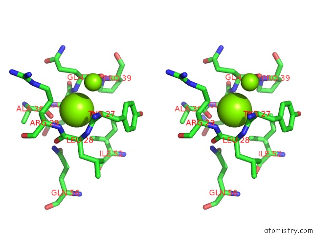
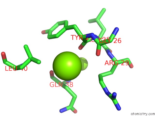
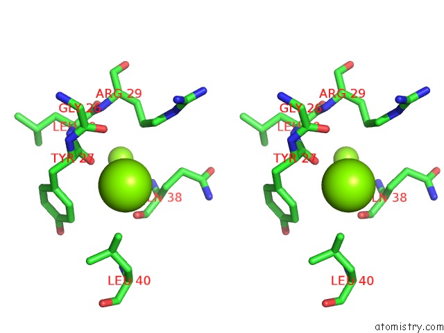
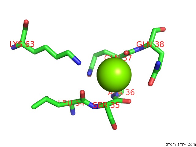
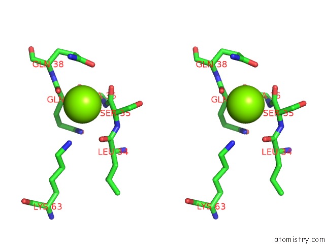
Magnesium binding site 1 out of 8 in 1yw0
Go back to
Magnesium binding site 1 out
of 8 in the Crystal Structure of the Tryptophan 2,3-Dioxygenase From Xanthomonas Campestris. Northeast Structural Genomics Target XCR13.
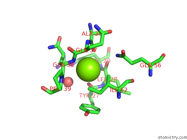
Mono view
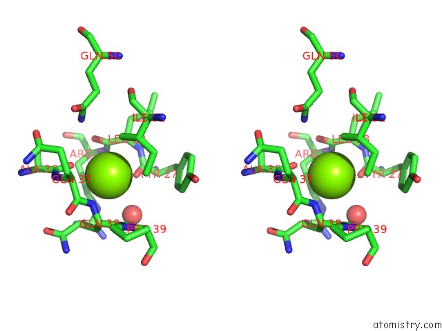
Stereo pair view
Mono view
Stereo pair view
A full contact list of Magnesium with other atoms in the Mg binding
site number 1 of Crystal Structure of the Tryptophan 2,3-Dioxygenase From Xanthomonas Campestris. Northeast Structural Genomics Target XCR13. within 5.0Å range:
|
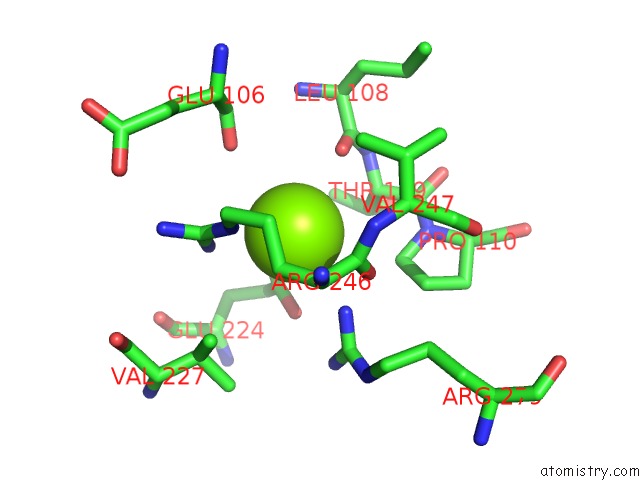
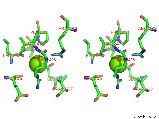
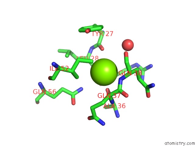
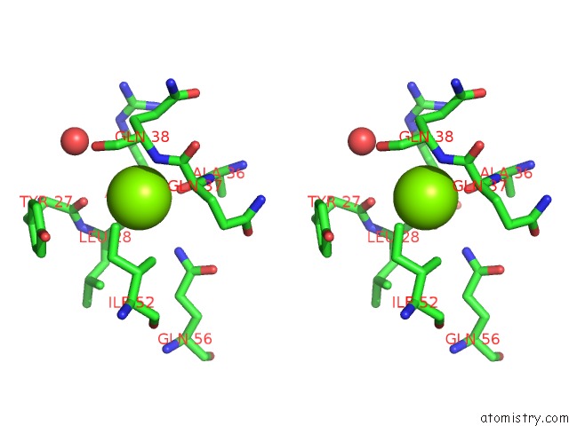
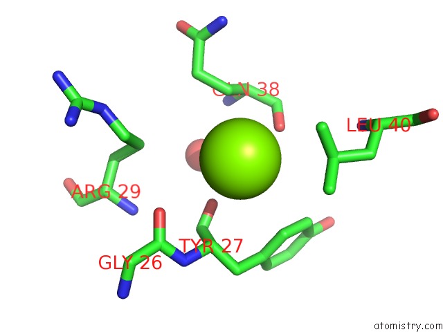
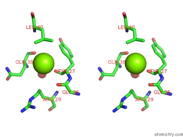
Magnesium binding site 2 out of 8 in 1yw0
Go back to
Magnesium binding site 2 out
of 8 in the Crystal Structure of the Tryptophan 2,3-Dioxygenase From Xanthomonas Campestris. Northeast Structural Genomics Target XCR13.
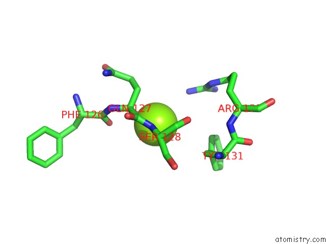
Mono view
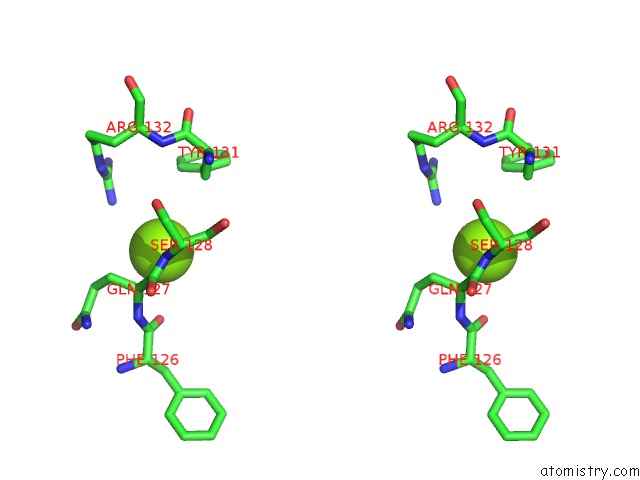
Stereo pair view
Mono view
Stereo pair view
A full contact list of Magnesium with other atoms in the Mg binding
site number 2 of Crystal Structure of the Tryptophan 2,3-Dioxygenase From Xanthomonas Campestris. Northeast Structural Genomics Target XCR13. within 5.0Å range:
|
Magnesium binding site 3 out of 8 in 1yw0
Go back to
Magnesium binding site 3 out
of 8 in the Crystal Structure of the Tryptophan 2,3-Dioxygenase From Xanthomonas Campestris. Northeast Structural Genomics Target XCR13.
Mono view
Stereo pair view
Mono view
Stereo pair view
A full contact list of Magnesium with other atoms in the Mg binding
site number 3 of Crystal Structure of the Tryptophan 2,3-Dioxygenase From Xanthomonas Campestris. Northeast Structural Genomics Target XCR13. within 5.0Å range:
|
Magnesium binding site 4 out of 8 in 1yw0
Go back to
Magnesium binding site 4 out
of 8 in the Crystal Structure of the Tryptophan 2,3-Dioxygenase From Xanthomonas Campestris. Northeast Structural Genomics Target XCR13.
Mono view
Stereo pair view
Mono view
Stereo pair view
A full contact list of Magnesium with other atoms in the Mg binding
site number 4 of Crystal Structure of the Tryptophan 2,3-Dioxygenase From Xanthomonas Campestris. Northeast Structural Genomics Target XCR13. within 5.0Å range:
|
Magnesium binding site 5 out of 8 in 1yw0
Go back to
Magnesium binding site 5 out
of 8 in the Crystal Structure of the Tryptophan 2,3-Dioxygenase From Xanthomonas Campestris. Northeast Structural Genomics Target XCR13.
Mono view
Stereo pair view
Mono view
Stereo pair view
A full contact list of Magnesium with other atoms in the Mg binding
site number 5 of Crystal Structure of the Tryptophan 2,3-Dioxygenase From Xanthomonas Campestris. Northeast Structural Genomics Target XCR13. within 5.0Å range:
|
Magnesium binding site 6 out of 8 in 1yw0
Go back to
Magnesium binding site 6 out
of 8 in the Crystal Structure of the Tryptophan 2,3-Dioxygenase From Xanthomonas Campestris. Northeast Structural Genomics Target XCR13.
Mono view
Stereo pair view
Mono view
Stereo pair view
A full contact list of Magnesium with other atoms in the Mg binding
site number 6 of Crystal Structure of the Tryptophan 2,3-Dioxygenase From Xanthomonas Campestris. Northeast Structural Genomics Target XCR13. within 5.0Å range:
|
Magnesium binding site 7 out of 8 in 1yw0
Go back to
Magnesium binding site 7 out
of 8 in the Crystal Structure of the Tryptophan 2,3-Dioxygenase From Xanthomonas Campestris. Northeast Structural Genomics Target XCR13.
Mono view
Stereo pair view
Mono view
Stereo pair view
A full contact list of Magnesium with other atoms in the Mg binding
site number 7 of Crystal Structure of the Tryptophan 2,3-Dioxygenase From Xanthomonas Campestris. Northeast Structural Genomics Target XCR13. within 5.0Å range:
|
Magnesium binding site 8 out of 8 in 1yw0
Go back to
Magnesium binding site 8 out
of 8 in the Crystal Structure of the Tryptophan 2,3-Dioxygenase From Xanthomonas Campestris. Northeast Structural Genomics Target XCR13.
Mono view
Stereo pair view
Mono view
Stereo pair view
A full contact list of Magnesium with other atoms in the Mg binding
site number 8 of Crystal Structure of the Tryptophan 2,3-Dioxygenase From Xanthomonas Campestris. Northeast Structural Genomics Target XCR13. within 5.0Å range:
|
Reference:
S.M.Vorobiev,
M.Abashidze,
F.Forouhar,
A.Kuzin,
R.Xiao,
M.Ciano,
T.B.Aton,
G.T.Montelione,
J.F.Hunt,
L.Tong.
Crystal Structure of the Tryptophan 2,3-Dioxygenase From Xanthomonas Campestris. Northeast Structural Genomics Target XCR13. To Be Published.
Page generated: Tue Aug 13 19:55:31 2024
Last articles
Zn in 9MJ5Zn in 9HNW
Zn in 9G0L
Zn in 9FNE
Zn in 9DZN
Zn in 9E0I
Zn in 9D32
Zn in 9DAK
Zn in 8ZXC
Zn in 8ZUF