Magnesium »
PDB 1yqz-1yzt »
1yxq »
Magnesium in PDB 1yxq: Crystal Structure of Actin in Complex with Swinholide A
Protein crystallography data
The structure of Crystal Structure of Actin in Complex with Swinholide A, PDB code: 1yxq
was solved by
V.A.Klenchin,
R.King,
J.Tanaka,
G.Marriott,
I.Rayment,
with X-Ray Crystallography technique. A brief refinement statistics is given in the table below:
| Resolution Low / High (Å) | 50.00 / 2.01 |
| Space group | P 1 21 1 |
| Cell size a, b, c (Å), α, β, γ (°) | 68.000, 76.800, 98.400, 90.00, 101.20, 90.00 |
| R / Rfree (%) | 18.4 / 21.9 |
Magnesium Binding Sites:
The binding sites of Magnesium atom in the Crystal Structure of Actin in Complex with Swinholide A
(pdb code 1yxq). This binding sites where shown within
5.0 Angstroms radius around Magnesium atom.
In total 3 binding sites of Magnesium where determined in the Crystal Structure of Actin in Complex with Swinholide A, PDB code: 1yxq:
Jump to Magnesium binding site number: 1; 2; 3;
In total 3 binding sites of Magnesium where determined in the Crystal Structure of Actin in Complex with Swinholide A, PDB code: 1yxq:
Jump to Magnesium binding site number: 1; 2; 3;
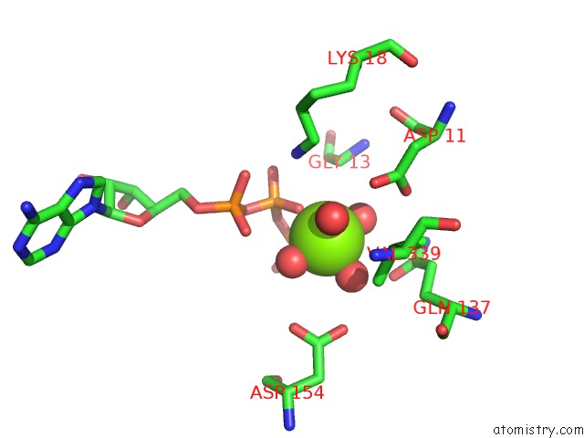
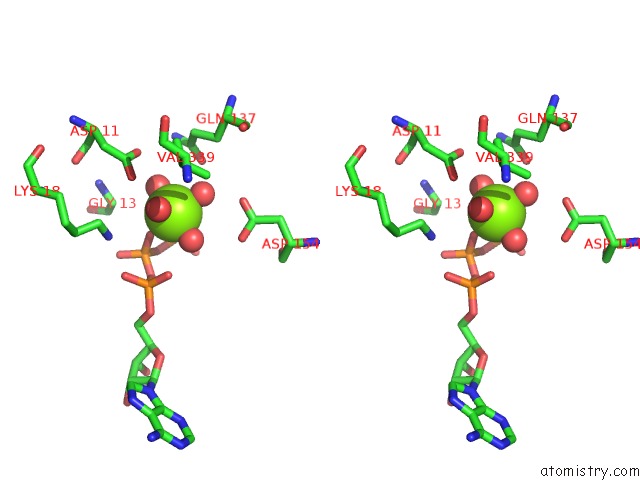
Magnesium binding site 1 out of 3 in 1yxq
Go back to
Magnesium binding site 1 out
of 3 in the Crystal Structure of Actin in Complex with Swinholide A
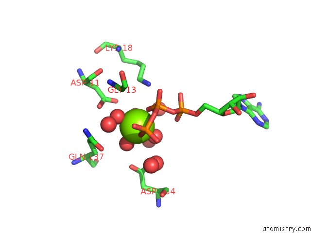
Mono view
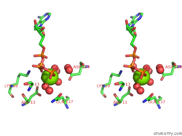
Stereo pair view
Mono view
Stereo pair view
A full contact list of Magnesium with other atoms in the Mg binding
site number 1 of Crystal Structure of Actin in Complex with Swinholide A within 5.0Å range:
|
Magnesium binding site 2 out of 3 in 1yxq
Go back to
Magnesium binding site 2 out
of 3 in the Crystal Structure of Actin in Complex with Swinholide A
Mono view
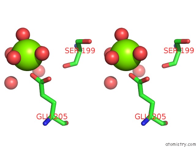
Stereo pair view
Mono view
Stereo pair view
A full contact list of Magnesium with other atoms in the Mg binding
site number 2 of Crystal Structure of Actin in Complex with Swinholide A within 5.0Å range:
|
Magnesium binding site 3 out of 3 in 1yxq
Go back to
Magnesium binding site 3 out
of 3 in the Crystal Structure of Actin in Complex with Swinholide A
Mono view
Stereo pair view
Mono view
Stereo pair view
A full contact list of Magnesium with other atoms in the Mg binding
site number 3 of Crystal Structure of Actin in Complex with Swinholide A within 5.0Å range:
|
Reference:
V.A.Klenchin,
R.King,
J.Tanaka,
G.Marriott,
I.Rayment.
Structural Basis of Swinholide A Binding to Actin Chem.Biol. V. 12 287 2005.
ISSN: ISSN 1074-5521
PubMed: 15797212
DOI: 10.1016/J.CHEMBIOL.2005.02.011
Page generated: Tue Aug 13 19:56:24 2024
ISSN: ISSN 1074-5521
PubMed: 15797212
DOI: 10.1016/J.CHEMBIOL.2005.02.011
Last articles
Zn in 9J0NZn in 9J0O
Zn in 9J0P
Zn in 9FJX
Zn in 9EKB
Zn in 9C0F
Zn in 9CAH
Zn in 9CH0
Zn in 9CH3
Zn in 9CH1