Magnesium »
PDB 208d-2a6e »
2a43 »
Magnesium in PDB 2a43: Crystal Structure of A Luteoviral Rna Pseudoknot and Model For A Minimal Ribosomal Frameshifting Motif
Protein crystallography data
The structure of Crystal Structure of A Luteoviral Rna Pseudoknot and Model For A Minimal Ribosomal Frameshifting Motif, PDB code: 2a43
was solved by
P.S.Pallan,
W.S.Marshall,
J.Harp,
F.C.Jewett Iii,
Z.Wawrzak,
B.A.Brown Ii,
A.Rich,
M.Egli,
with X-Ray Crystallography technique. A brief refinement statistics is given in the table below:
| Resolution Low / High (Å) | 23.60 / 1.34 |
| Space group | P 32 2 1 |
| Cell size a, b, c (Å), α, β, γ (°) | 53.250, 53.250, 55.000, 90.00, 90.00, 120.00 |
| R / Rfree (%) | n/a / n/a |
Magnesium Binding Sites:
The binding sites of Magnesium atom in the Crystal Structure of A Luteoviral Rna Pseudoknot and Model For A Minimal Ribosomal Frameshifting Motif
(pdb code 2a43). This binding sites where shown within
5.0 Angstroms radius around Magnesium atom.
In total 2 binding sites of Magnesium where determined in the Crystal Structure of A Luteoviral Rna Pseudoknot and Model For A Minimal Ribosomal Frameshifting Motif, PDB code: 2a43:
Jump to Magnesium binding site number: 1; 2;
In total 2 binding sites of Magnesium where determined in the Crystal Structure of A Luteoviral Rna Pseudoknot and Model For A Minimal Ribosomal Frameshifting Motif, PDB code: 2a43:
Jump to Magnesium binding site number: 1; 2;
Magnesium binding site 1 out of 2 in 2a43
Go back to
Magnesium binding site 1 out
of 2 in the Crystal Structure of A Luteoviral Rna Pseudoknot and Model For A Minimal Ribosomal Frameshifting Motif
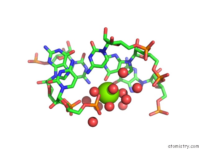
Mono view
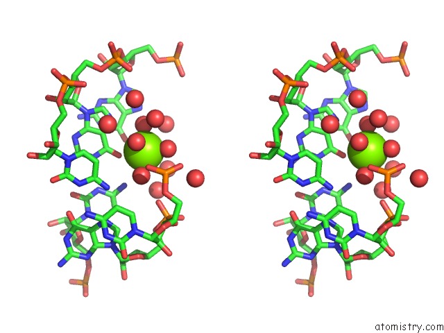
Stereo pair view
Mono view
Stereo pair view
A full contact list of Magnesium with other atoms in the Mg binding
site number 1 of Crystal Structure of A Luteoviral Rna Pseudoknot and Model For A Minimal Ribosomal Frameshifting Motif within 5.0Å range:
|
Magnesium binding site 2 out of 2 in 2a43
Go back to
Magnesium binding site 2 out
of 2 in the Crystal Structure of A Luteoviral Rna Pseudoknot and Model For A Minimal Ribosomal Frameshifting Motif

Mono view
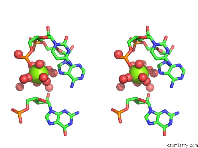
Stereo pair view
Mono view
Stereo pair view
A full contact list of Magnesium with other atoms in the Mg binding
site number 2 of Crystal Structure of A Luteoviral Rna Pseudoknot and Model For A Minimal Ribosomal Frameshifting Motif within 5.0Å range:
|
Reference:
P.S.Pallan,
W.S.Marshall,
J.Harp,
F.C.Jewett Iii,
Z.Wawrzak,
B.A.Brown Ii,
A.Rich,
M.Egli.
Crystal Structure of A Luteoviral Rna Pseudoknot and Model For A Minimal Ribosomal Frameshifting Motif Biochemistry V. 44 11315 2005.
ISSN: ISSN 0006-2960
PubMed: 16114868
DOI: 10.1021/BI051061I
Page generated: Tue Aug 13 20:22:27 2024
ISSN: ISSN 0006-2960
PubMed: 16114868
DOI: 10.1021/BI051061I
Last articles
Fe in 2YXOFe in 2YRS
Fe in 2YXC
Fe in 2YNM
Fe in 2YVJ
Fe in 2YP1
Fe in 2YU2
Fe in 2YU1
Fe in 2YQB
Fe in 2YOO