Magnesium »
PDB 2a6h-2alz »
2a87 »
Magnesium in PDB 2a87: Crystal Structure of M. Tuberculosis Thioredoxin Reductase
Enzymatic activity of Crystal Structure of M. Tuberculosis Thioredoxin Reductase
All present enzymatic activity of Crystal Structure of M. Tuberculosis Thioredoxin Reductase:
1.8.1.9;
1.8.1.9;
Protein crystallography data
The structure of Crystal Structure of M. Tuberculosis Thioredoxin Reductase, PDB code: 2a87
was solved by
M.Akif,
K.Suhre,
C.Verma,
S.C.Mande,
Tb Structural Genomics Consortium(Tbsgc),
with X-Ray Crystallography technique. A brief refinement statistics is given in the table below:
| Resolution Low / High (Å) | 36.00 / 3.00 |
| Space group | P 41 21 2 |
| Cell size a, b, c (Å), α, β, γ (°) | 107.400, 107.400, 118.200, 90.00, 90.00, 90.00 |
| R / Rfree (%) | 21.1 / 29.1 |
Magnesium Binding Sites:
The binding sites of Magnesium atom in the Crystal Structure of M. Tuberculosis Thioredoxin Reductase
(pdb code 2a87). This binding sites where shown within
5.0 Angstroms radius around Magnesium atom.
In total 2 binding sites of Magnesium where determined in the Crystal Structure of M. Tuberculosis Thioredoxin Reductase, PDB code: 2a87:
Jump to Magnesium binding site number: 1; 2;
In total 2 binding sites of Magnesium where determined in the Crystal Structure of M. Tuberculosis Thioredoxin Reductase, PDB code: 2a87:
Jump to Magnesium binding site number: 1; 2;
Magnesium binding site 1 out of 2 in 2a87
Go back to
Magnesium binding site 1 out
of 2 in the Crystal Structure of M. Tuberculosis Thioredoxin Reductase
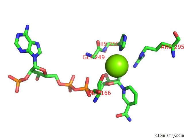
Mono view
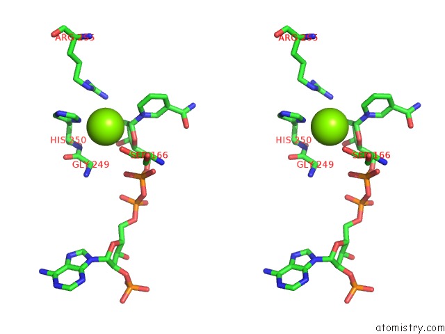
Stereo pair view
Mono view
Stereo pair view
A full contact list of Magnesium with other atoms in the Mg binding
site number 1 of Crystal Structure of M. Tuberculosis Thioredoxin Reductase within 5.0Å range:
|
Magnesium binding site 2 out of 2 in 2a87
Go back to
Magnesium binding site 2 out
of 2 in the Crystal Structure of M. Tuberculosis Thioredoxin Reductase
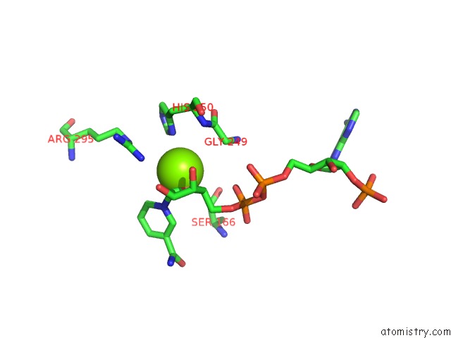
Mono view
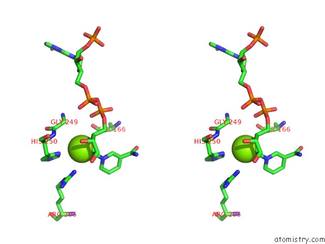
Stereo pair view
Mono view
Stereo pair view
A full contact list of Magnesium with other atoms in the Mg binding
site number 2 of Crystal Structure of M. Tuberculosis Thioredoxin Reductase within 5.0Å range:
|
Reference:
M.Akif,
K.Suhre,
C.Verma,
S.C.Mande.
Conformational Flexibility of Mycobacterium Tuberculosis Thioredoxin Reductase: Crystal Structure and Normal-Mode Analysis. Acta Crystallogr.,Sect.D V. 61 1603 2005.
ISSN: ISSN 0907-4449
PubMed: 16301794
DOI: 10.1107/S0907444905030519
Page generated: Sun Aug 10 09:43:18 2025
ISSN: ISSN 0907-4449
PubMed: 16301794
DOI: 10.1107/S0907444905030519
Last articles
Mg in 6CA4Mg in 6C90
Mg in 6CA0
Mg in 6C9Y
Mg in 6C8Z
Mg in 6C8P
Mg in 6C8N
Mg in 6C8O
Mg in 6C8D
Mg in 6C8L