Magnesium »
PDB 2a6h-2alz »
2acx »
Magnesium in PDB 2acx: Crystal Structure of G Protein Coupled Receptor Kinase 6 Bound to Amppnp
Protein crystallography data
The structure of Crystal Structure of G Protein Coupled Receptor Kinase 6 Bound to Amppnp, PDB code: 2acx
was solved by
D.T.Lodowski,
V.M.Tesmer,
J.L.Benovic,
J.J.Tesmer,
with X-Ray Crystallography technique. A brief refinement statistics is given in the table below:
| Resolution Low / High (Å) | 30.00 / 2.60 |
| Space group | C 1 2 1 |
| Cell size a, b, c (Å), α, β, γ (°) | 120.164, 59.270, 221.095, 90.00, 102.58, 90.00 |
| R / Rfree (%) | 20.2 / 24.3 |
Magnesium Binding Sites:
The binding sites of Magnesium atom in the Crystal Structure of G Protein Coupled Receptor Kinase 6 Bound to Amppnp
(pdb code 2acx). This binding sites where shown within
5.0 Angstroms radius around Magnesium atom.
In total 2 binding sites of Magnesium where determined in the Crystal Structure of G Protein Coupled Receptor Kinase 6 Bound to Amppnp, PDB code: 2acx:
Jump to Magnesium binding site number: 1; 2;
In total 2 binding sites of Magnesium where determined in the Crystal Structure of G Protein Coupled Receptor Kinase 6 Bound to Amppnp, PDB code: 2acx:
Jump to Magnesium binding site number: 1; 2;
Magnesium binding site 1 out of 2 in 2acx
Go back to
Magnesium binding site 1 out
of 2 in the Crystal Structure of G Protein Coupled Receptor Kinase 6 Bound to Amppnp
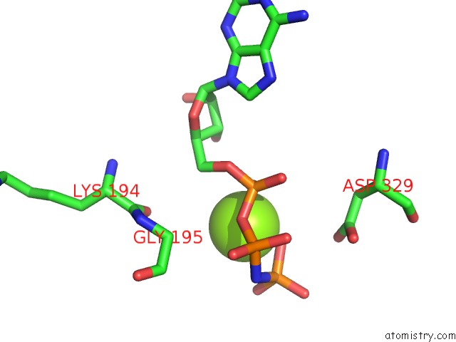
Mono view
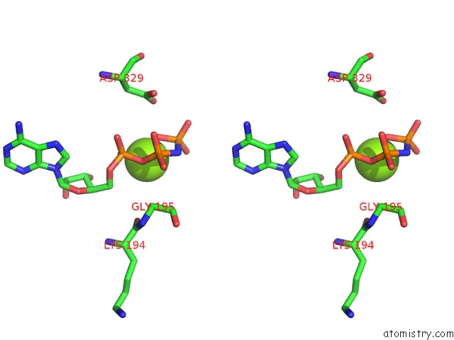
Stereo pair view
Mono view
Stereo pair view
A full contact list of Magnesium with other atoms in the Mg binding
site number 1 of Crystal Structure of G Protein Coupled Receptor Kinase 6 Bound to Amppnp within 5.0Å range:
|
Magnesium binding site 2 out of 2 in 2acx
Go back to
Magnesium binding site 2 out
of 2 in the Crystal Structure of G Protein Coupled Receptor Kinase 6 Bound to Amppnp
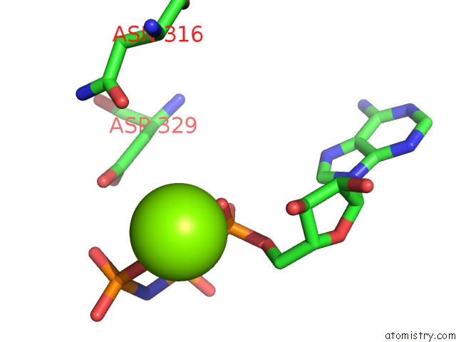
Mono view
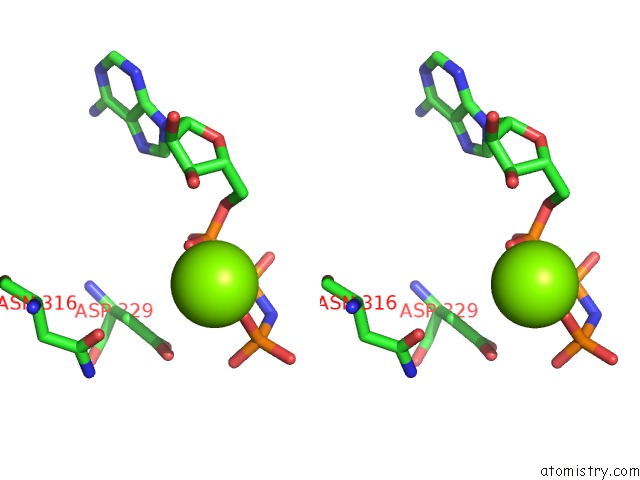
Stereo pair view
Mono view
Stereo pair view
A full contact list of Magnesium with other atoms in the Mg binding
site number 2 of Crystal Structure of G Protein Coupled Receptor Kinase 6 Bound to Amppnp within 5.0Å range:
|
Reference:
D.T.Lodowski,
V.M.Tesmer,
J.L.Benovic,
J.J.Tesmer.
The Structure of G Protein-Coupled Receptor Kinase (Grk)-6 Defines A Second Lineage of Grks. J.Biol.Chem. V. 281 16785 2006.
ISSN: ISSN 0021-9258
PubMed: 16613860
DOI: 10.1074/JBC.M601327200
Page generated: Sun Aug 10 09:43:39 2025
ISSN: ISSN 0021-9258
PubMed: 16613860
DOI: 10.1074/JBC.M601327200
Last articles
Mg in 6LU1Mg in 6LT4
Mg in 6LY7
Mg in 6LY6
Mg in 6LY3
Mg in 6LX1
Mg in 6LTW
Mg in 6LVW
Mg in 6LUH
Mg in 6LTS