Magnesium »
PDB 2amc-2b2k »
2amc »
Magnesium in PDB 2amc: Crystal Structure of Phenylalanyl-Trna Synthetase Complexed with L- Tyrosine
Enzymatic activity of Crystal Structure of Phenylalanyl-Trna Synthetase Complexed with L- Tyrosine
All present enzymatic activity of Crystal Structure of Phenylalanyl-Trna Synthetase Complexed with L- Tyrosine:
6.1.1.20;
6.1.1.20;
Protein crystallography data
The structure of Crystal Structure of Phenylalanyl-Trna Synthetase Complexed with L- Tyrosine, PDB code: 2amc
was solved by
O.Kotik-Kogan,
N.Moor,
D.Tworowski,
M.Safro,
with X-Ray Crystallography technique. A brief refinement statistics is given in the table below:
| Resolution Low / High (Å) | 25.41 / 2.70 |
| Space group | P 32 2 1 |
| Cell size a, b, c (Å), α, β, γ (°) | 173.200, 173.200, 138.230, 90.00, 90.00, 120.00 |
| R / Rfree (%) | 22.9 / 25.5 |
Magnesium Binding Sites:
The binding sites of Magnesium atom in the Crystal Structure of Phenylalanyl-Trna Synthetase Complexed with L- Tyrosine
(pdb code 2amc). This binding sites where shown within
5.0 Angstroms radius around Magnesium atom.
In total only one binding site of Magnesium was determined in the Crystal Structure of Phenylalanyl-Trna Synthetase Complexed with L- Tyrosine, PDB code: 2amc:
In total only one binding site of Magnesium was determined in the Crystal Structure of Phenylalanyl-Trna Synthetase Complexed with L- Tyrosine, PDB code: 2amc:
Magnesium binding site 1 out of 1 in 2amc
Go back to
Magnesium binding site 1 out
of 1 in the Crystal Structure of Phenylalanyl-Trna Synthetase Complexed with L- Tyrosine
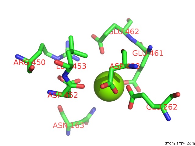
Mono view
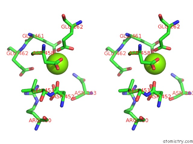
Stereo pair view
Mono view
Stereo pair view
A full contact list of Magnesium with other atoms in the Mg binding
site number 1 of Crystal Structure of Phenylalanyl-Trna Synthetase Complexed with L- Tyrosine within 5.0Å range:
|
Reference:
O.Kotik-Kogan,
N.Moor,
D.Tworowski,
M.Safro.
Structural Basis For Discrimination of L-Phenylalanine From L-Tyrosine By Phenylalanyl-Trna Synthetase Structure V. 13 1799 2005.
ISSN: ISSN 0969-2126
PubMed: 16338408
DOI: 10.1016/J.STR.2005.08.013
Page generated: Tue Aug 13 21:35:01 2024
ISSN: ISSN 0969-2126
PubMed: 16338408
DOI: 10.1016/J.STR.2005.08.013
Last articles
Zn in 9J0NZn in 9J0O
Zn in 9J0P
Zn in 9FJX
Zn in 9EKB
Zn in 9C0F
Zn in 9CAH
Zn in 9CH0
Zn in 9CH3
Zn in 9CH1