Magnesium »
PDB 2b2x-2bhc »
2b9f »
Magnesium in PDB 2b9f: Crystal Structure of Non-Phosphorylated FUS3
Enzymatic activity of Crystal Structure of Non-Phosphorylated FUS3
All present enzymatic activity of Crystal Structure of Non-Phosphorylated FUS3:
2.7.1.37;
2.7.1.37;
Protein crystallography data
The structure of Crystal Structure of Non-Phosphorylated FUS3, PDB code: 2b9f
was solved by
A.Remenyi,
M.C.Good,
R.P.Bhattacharyya,
W.A.Lim,
with X-Ray Crystallography technique. A brief refinement statistics is given in the table below:
| Resolution Low / High (Å) | 20.00 / 1.80 |
| Space group | P 21 21 21 |
| Cell size a, b, c (Å), α, β, γ (°) | 56.700, 62.470, 87.168, 90.00, 90.00, 90.00 |
| R / Rfree (%) | 21.3 / 25.9 |
Magnesium Binding Sites:
The binding sites of Magnesium atom in the Crystal Structure of Non-Phosphorylated FUS3
(pdb code 2b9f). This binding sites where shown within
5.0 Angstroms radius around Magnesium atom.
In total only one binding site of Magnesium was determined in the Crystal Structure of Non-Phosphorylated FUS3, PDB code: 2b9f:
In total only one binding site of Magnesium was determined in the Crystal Structure of Non-Phosphorylated FUS3, PDB code: 2b9f:
Magnesium binding site 1 out of 1 in 2b9f
Go back to
Magnesium binding site 1 out
of 1 in the Crystal Structure of Non-Phosphorylated FUS3
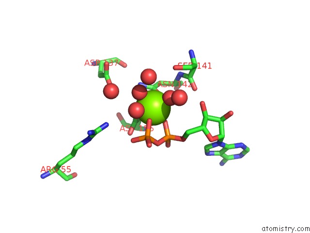
Mono view
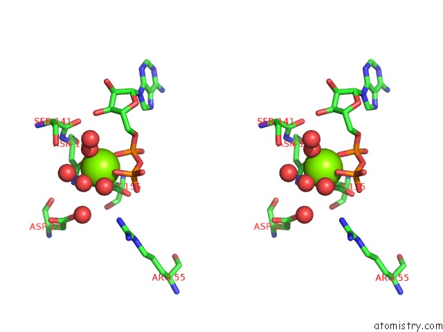
Stereo pair view
Mono view
Stereo pair view
A full contact list of Magnesium with other atoms in the Mg binding
site number 1 of Crystal Structure of Non-Phosphorylated FUS3 within 5.0Å range:
|
Reference:
A.Remenyi,
M.C.Good,
R.P.Bhattacharyya,
W.A.Lim.
The Role of Docking Interactions in Mediating Signaling Input, Output, and Discrimination in the Yeast Mapk Network. Mol.Cell V. 20 951 2005.
ISSN: ISSN 1097-2765
PubMed: 16364919
DOI: 10.1016/J.MOLCEL.2005.10.030
Page generated: Tue Aug 13 21:48:09 2024
ISSN: ISSN 1097-2765
PubMed: 16364919
DOI: 10.1016/J.MOLCEL.2005.10.030
Last articles
Zn in 9J0NZn in 9J0O
Zn in 9J0P
Zn in 9FJX
Zn in 9EKB
Zn in 9C0F
Zn in 9CAH
Zn in 9CH0
Zn in 9CH3
Zn in 9CH1