Magnesium »
PDB 2b2x-2bhc »
2bcr »
Magnesium in PDB 2bcr: Dna Polymerase Lambda in Complex with A Dna Duplex Containing An Unpaired Damp
Enzymatic activity of Dna Polymerase Lambda in Complex with A Dna Duplex Containing An Unpaired Damp
All present enzymatic activity of Dna Polymerase Lambda in Complex with A Dna Duplex Containing An Unpaired Damp:
2.7.7.7;
2.7.7.7;
Protein crystallography data
The structure of Dna Polymerase Lambda in Complex with A Dna Duplex Containing An Unpaired Damp, PDB code: 2bcr
was solved by
M.Garcia-Diaz,
K.Bebenek,
J.M.Krahn,
L.C.Pedersen,
T.A.Kunkel,
with X-Ray Crystallography technique. A brief refinement statistics is given in the table below:
| Resolution Low / High (Å) | 41.87 / 1.75 |
| Space group | P 21 21 21 |
| Cell size a, b, c (Å), α, β, γ (°) | 56.076, 62.925, 139.364, 90.00, 90.00, 90.00 |
| R / Rfree (%) | 20.2 / 23.1 |
Other elements in 2bcr:
The structure of Dna Polymerase Lambda in Complex with A Dna Duplex Containing An Unpaired Damp also contains other interesting chemical elements:
| Sodium | (Na) | 3 atoms |
Magnesium Binding Sites:
The binding sites of Magnesium atom in the Dna Polymerase Lambda in Complex with A Dna Duplex Containing An Unpaired Damp
(pdb code 2bcr). This binding sites where shown within
5.0 Angstroms radius around Magnesium atom.
In total only one binding site of Magnesium was determined in the Dna Polymerase Lambda in Complex with A Dna Duplex Containing An Unpaired Damp, PDB code: 2bcr:
In total only one binding site of Magnesium was determined in the Dna Polymerase Lambda in Complex with A Dna Duplex Containing An Unpaired Damp, PDB code: 2bcr:
Magnesium binding site 1 out of 1 in 2bcr
Go back to
Magnesium binding site 1 out
of 1 in the Dna Polymerase Lambda in Complex with A Dna Duplex Containing An Unpaired Damp
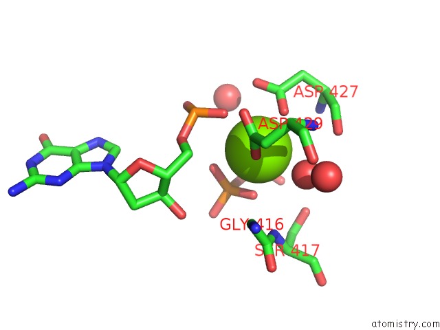
Mono view
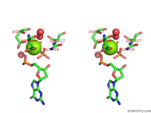
Stereo pair view
Mono view
Stereo pair view
A full contact list of Magnesium with other atoms in the Mg binding
site number 1 of Dna Polymerase Lambda in Complex with A Dna Duplex Containing An Unpaired Damp within 5.0Å range:
|
Reference:
M.Garcia-Diaz,
K.Bebenek,
J.M.Krahn,
L.C.Pedersen,
T.A.Kunkel.
Structural Analysis of Strand Misalignment During Dna Synthesis By A Human Dna Polymerase Cell(Cambridge,Mass.) V. 124 331 2006.
ISSN: ISSN 0092-8674
PubMed: 16439207
DOI: 10.1016/J.CELL.2005.10.039
Page generated: Tue Aug 13 21:50:02 2024
ISSN: ISSN 0092-8674
PubMed: 16439207
DOI: 10.1016/J.CELL.2005.10.039
Last articles
Zn in 9J0NZn in 9J0O
Zn in 9J0P
Zn in 9FJX
Zn in 9EKB
Zn in 9C0F
Zn in 9CAH
Zn in 9CH0
Zn in 9CH3
Zn in 9CH1