Magnesium »
PDB 2bhd-2bt1 »
2bhy »
Magnesium in PDB 2bhy: Crystal Structure of Deinococcus Radiodurans Maltooligosyltrehalose Trehalohydrolase in Complex with Trehalose
Enzymatic activity of Crystal Structure of Deinococcus Radiodurans Maltooligosyltrehalose Trehalohydrolase in Complex with Trehalose
All present enzymatic activity of Crystal Structure of Deinococcus Radiodurans Maltooligosyltrehalose Trehalohydrolase in Complex with Trehalose:
3.2.1.1;
3.2.1.1;
Protein crystallography data
The structure of Crystal Structure of Deinococcus Radiodurans Maltooligosyltrehalose Trehalohydrolase in Complex with Trehalose, PDB code: 2bhy
was solved by
J.Timmins,
H.-K.S.Leiros,
G.Leonard,
I.Leiros,
S.Mcsweeney,
with X-Ray Crystallography technique. A brief refinement statistics is given in the table below:
| Resolution Low / High (Å) | 76.70 / 1.5 |
| Space group | P 21 21 21 |
| Cell size a, b, c (Å), α, β, γ (°) | 60.029, 66.556, 153.141, 90.00, 90.00, 90.00 |
| R / Rfree (%) | 12.8 / 15.2 |
Magnesium Binding Sites:
The binding sites of Magnesium atom in the Crystal Structure of Deinococcus Radiodurans Maltooligosyltrehalose Trehalohydrolase in Complex with Trehalose
(pdb code 2bhy). This binding sites where shown within
5.0 Angstroms radius around Magnesium atom.
In total only one binding site of Magnesium was determined in the Crystal Structure of Deinococcus Radiodurans Maltooligosyltrehalose Trehalohydrolase in Complex with Trehalose, PDB code: 2bhy:
In total only one binding site of Magnesium was determined in the Crystal Structure of Deinococcus Radiodurans Maltooligosyltrehalose Trehalohydrolase in Complex with Trehalose, PDB code: 2bhy:
Magnesium binding site 1 out of 1 in 2bhy
Go back to
Magnesium binding site 1 out
of 1 in the Crystal Structure of Deinococcus Radiodurans Maltooligosyltrehalose Trehalohydrolase in Complex with Trehalose
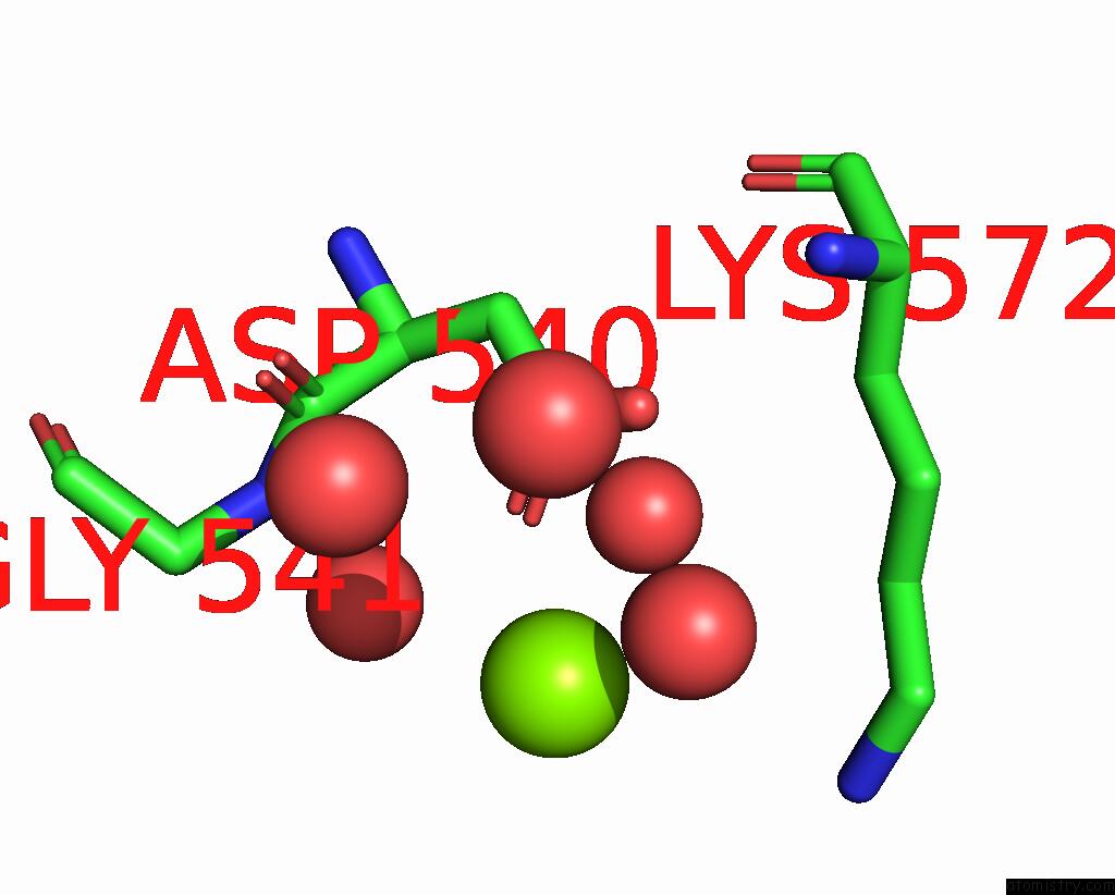
Mono view
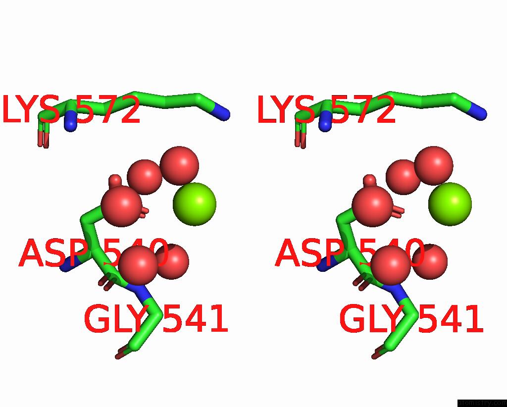
Stereo pair view
Mono view
Stereo pair view
A full contact list of Magnesium with other atoms in the Mg binding
site number 1 of Crystal Structure of Deinococcus Radiodurans Maltooligosyltrehalose Trehalohydrolase in Complex with Trehalose within 5.0Å range:
|
Reference:
J.Timmins,
H.-K.S.Leiros,
G.Leonard,
I.Leiros,
S.Mcsweeney.
Crystal Structure of Maltooligosyltrehalose Trehalohydrolase From Deinococcus Radiodurans in Complex with Disaccharides J.Mol.Biol. V. 347 949 2005.
ISSN: ISSN 0022-2836
PubMed: 15784255
DOI: 10.1016/J.JMB.2005.02.011
Page generated: Sun Aug 10 10:00:04 2025
ISSN: ISSN 0022-2836
PubMed: 15784255
DOI: 10.1016/J.JMB.2005.02.011
Last articles
Mg in 5SI5Mg in 5SI4
Mg in 5SI3
Mg in 5SI2
Mg in 5SI0
Mg in 5SHZ
Mg in 5SI1
Mg in 5SHX
Mg in 5SHW
Mg in 5SHY