Magnesium »
PDB 2bhd-2bt1 »
2bie »
Magnesium in PDB 2bie: Radiation Damage of the Schiff Base in Phosphoserine Aminotransferase (Structure H)
Enzymatic activity of Radiation Damage of the Schiff Base in Phosphoserine Aminotransferase (Structure H)
All present enzymatic activity of Radiation Damage of the Schiff Base in Phosphoserine Aminotransferase (Structure H):
2.6.1.52;
2.6.1.52;
Protein crystallography data
The structure of Radiation Damage of the Schiff Base in Phosphoserine Aminotransferase (Structure H), PDB code: 2bie
was solved by
A.P.Dubnovitsky,
R.B.G.Ravelli,
A.N.Popov,
A.C.Papageorgiou,
with X-Ray Crystallography technique. A brief refinement statistics is given in the table below:
| Resolution Low / High (Å) | 12.00 / 1.30 |
| Space group | P 21 21 2 |
| Cell size a, b, c (Å), α, β, γ (°) | 143.730, 84.271, 67.207, 90.00, 90.00, 90.00 |
| R / Rfree (%) | 12.9 / 16.4 |
Other elements in 2bie:
The structure of Radiation Damage of the Schiff Base in Phosphoserine Aminotransferase (Structure H) also contains other interesting chemical elements:
| Chlorine | (Cl) | 4 atoms |
Magnesium Binding Sites:
The binding sites of Magnesium atom in the Radiation Damage of the Schiff Base in Phosphoserine Aminotransferase (Structure H)
(pdb code 2bie). This binding sites where shown within
5.0 Angstroms radius around Magnesium atom.
In total 6 binding sites of Magnesium where determined in the Radiation Damage of the Schiff Base in Phosphoserine Aminotransferase (Structure H), PDB code: 2bie:
Jump to Magnesium binding site number: 1; 2; 3; 4; 5; 6;
In total 6 binding sites of Magnesium where determined in the Radiation Damage of the Schiff Base in Phosphoserine Aminotransferase (Structure H), PDB code: 2bie:
Jump to Magnesium binding site number: 1; 2; 3; 4; 5; 6;
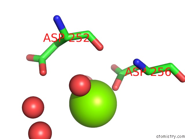
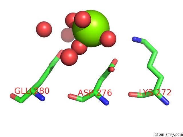
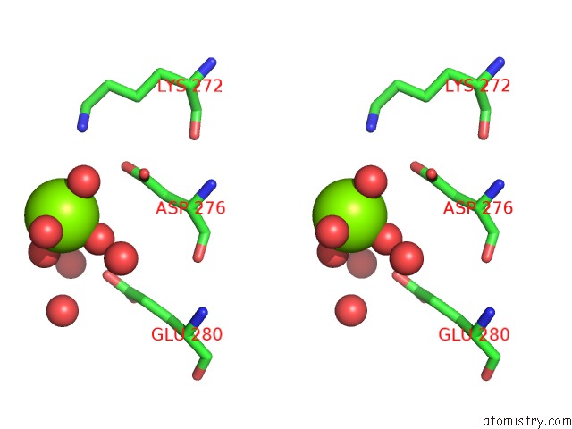
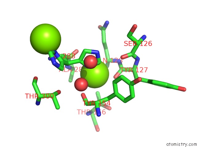
Magnesium binding site 1 out of 6 in 2bie
Go back to
Magnesium binding site 1 out
of 6 in the Radiation Damage of the Schiff Base in Phosphoserine Aminotransferase (Structure H)
Mono view
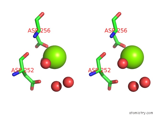
Stereo pair view
Mono view
Stereo pair view
A full contact list of Magnesium with other atoms in the Mg binding
site number 1 of Radiation Damage of the Schiff Base in Phosphoserine Aminotransferase (Structure H) within 5.0Å range:
|
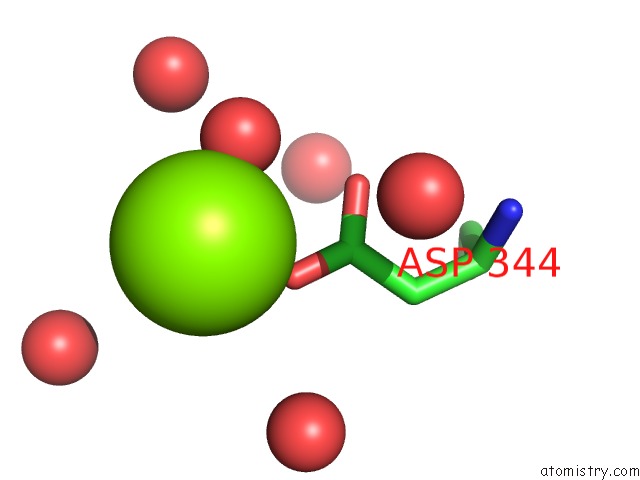
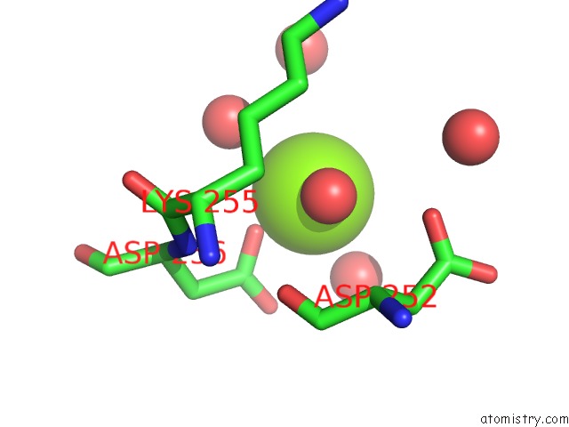
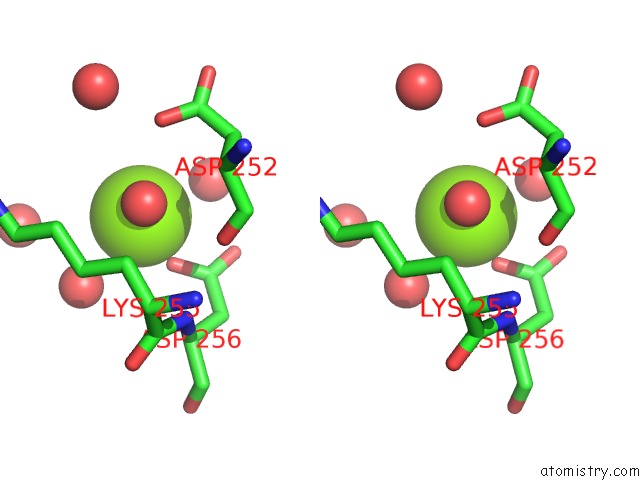
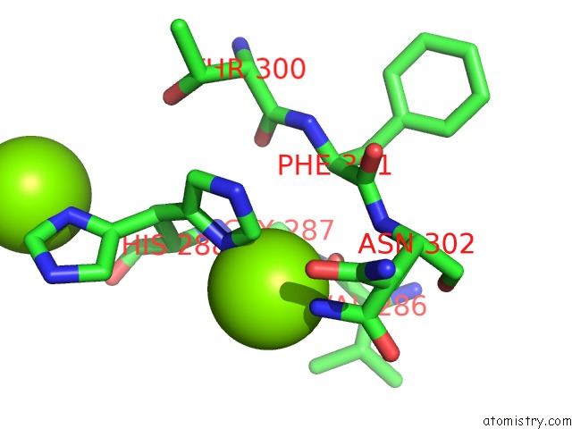
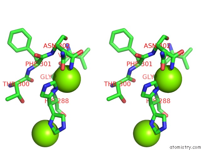
Magnesium binding site 2 out of 6 in 2bie
Go back to
Magnesium binding site 2 out
of 6 in the Radiation Damage of the Schiff Base in Phosphoserine Aminotransferase (Structure H)
Mono view
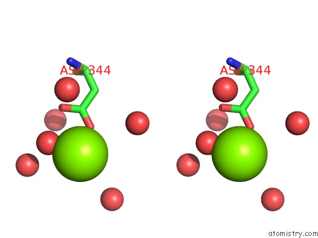
Stereo pair view
Mono view
Stereo pair view
A full contact list of Magnesium with other atoms in the Mg binding
site number 2 of Radiation Damage of the Schiff Base in Phosphoserine Aminotransferase (Structure H) within 5.0Å range:
|
Magnesium binding site 3 out of 6 in 2bie
Go back to
Magnesium binding site 3 out
of 6 in the Radiation Damage of the Schiff Base in Phosphoserine Aminotransferase (Structure H)
Mono view
Stereo pair view
Mono view
Stereo pair view
A full contact list of Magnesium with other atoms in the Mg binding
site number 3 of Radiation Damage of the Schiff Base in Phosphoserine Aminotransferase (Structure H) within 5.0Å range:
|
Magnesium binding site 4 out of 6 in 2bie
Go back to
Magnesium binding site 4 out
of 6 in the Radiation Damage of the Schiff Base in Phosphoserine Aminotransferase (Structure H)
Mono view
Stereo pair view
Mono view
Stereo pair view
A full contact list of Magnesium with other atoms in the Mg binding
site number 4 of Radiation Damage of the Schiff Base in Phosphoserine Aminotransferase (Structure H) within 5.0Å range:
|
Magnesium binding site 5 out of 6 in 2bie
Go back to
Magnesium binding site 5 out
of 6 in the Radiation Damage of the Schiff Base in Phosphoserine Aminotransferase (Structure H)
Mono view

Stereo pair view
Mono view
Stereo pair view
A full contact list of Magnesium with other atoms in the Mg binding
site number 5 of Radiation Damage of the Schiff Base in Phosphoserine Aminotransferase (Structure H) within 5.0Å range:
|
Magnesium binding site 6 out of 6 in 2bie
Go back to
Magnesium binding site 6 out
of 6 in the Radiation Damage of the Schiff Base in Phosphoserine Aminotransferase (Structure H)
Mono view
Stereo pair view
Mono view
Stereo pair view
A full contact list of Magnesium with other atoms in the Mg binding
site number 6 of Radiation Damage of the Schiff Base in Phosphoserine Aminotransferase (Structure H) within 5.0Å range:
|
Reference:
A.P.Dubnovitsky,
R.B.G.Ravelli,
A.N.Popov,
A.C.Papageorgiou.
Strain Relief at the Active Site of Phosphoserine Aminotransferase Induced By Radiation Damage. Protein Sci. V. 14 1498 2005.
ISSN: ISSN 0961-8368
PubMed: 15883191
DOI: 10.1110/PS.051397905
Page generated: Sun Aug 10 10:02:00 2025
ISSN: ISSN 0961-8368
PubMed: 15883191
DOI: 10.1110/PS.051397905
Last articles
Mg in 2WTZMg in 2WTY
Mg in 2WTP
Mg in 2WTO
Mg in 2WSS
Mg in 2WSB
Mg in 2WPD
Mg in 2WOQ
Mg in 2WOJ
Mg in 2WQS