Magnesium »
PDB 2bhd-2bt1 »
2bon »
Magnesium in PDB 2bon: Structure of An Escherichia Coli Lipid Kinase (Yegs)
Protein crystallography data
The structure of Structure of An Escherichia Coli Lipid Kinase (Yegs), PDB code: 2bon
was solved by
H.M.Bakali,
K.A.Johnson,
B.M.Hallberg,
M.D.Herman,
P.Nordlund,
with X-Ray Crystallography technique. A brief refinement statistics is given in the table below:
| Resolution Low / High (Å) | 83.04 / 1.90 |
| Space group | P 1 21 1 |
| Cell size a, b, c (Å), α, β, γ (°) | 42.326, 166.164, 48.474, 90.00, 97.03, 90.00 |
| R / Rfree (%) | 22 / 26.7 |
Magnesium Binding Sites:
The binding sites of Magnesium atom in the Structure of An Escherichia Coli Lipid Kinase (Yegs)
(pdb code 2bon). This binding sites where shown within
5.0 Angstroms radius around Magnesium atom.
In total 2 binding sites of Magnesium where determined in the Structure of An Escherichia Coli Lipid Kinase (Yegs), PDB code: 2bon:
Jump to Magnesium binding site number: 1; 2;
In total 2 binding sites of Magnesium where determined in the Structure of An Escherichia Coli Lipid Kinase (Yegs), PDB code: 2bon:
Jump to Magnesium binding site number: 1; 2;
Magnesium binding site 1 out of 2 in 2bon
Go back to
Magnesium binding site 1 out
of 2 in the Structure of An Escherichia Coli Lipid Kinase (Yegs)
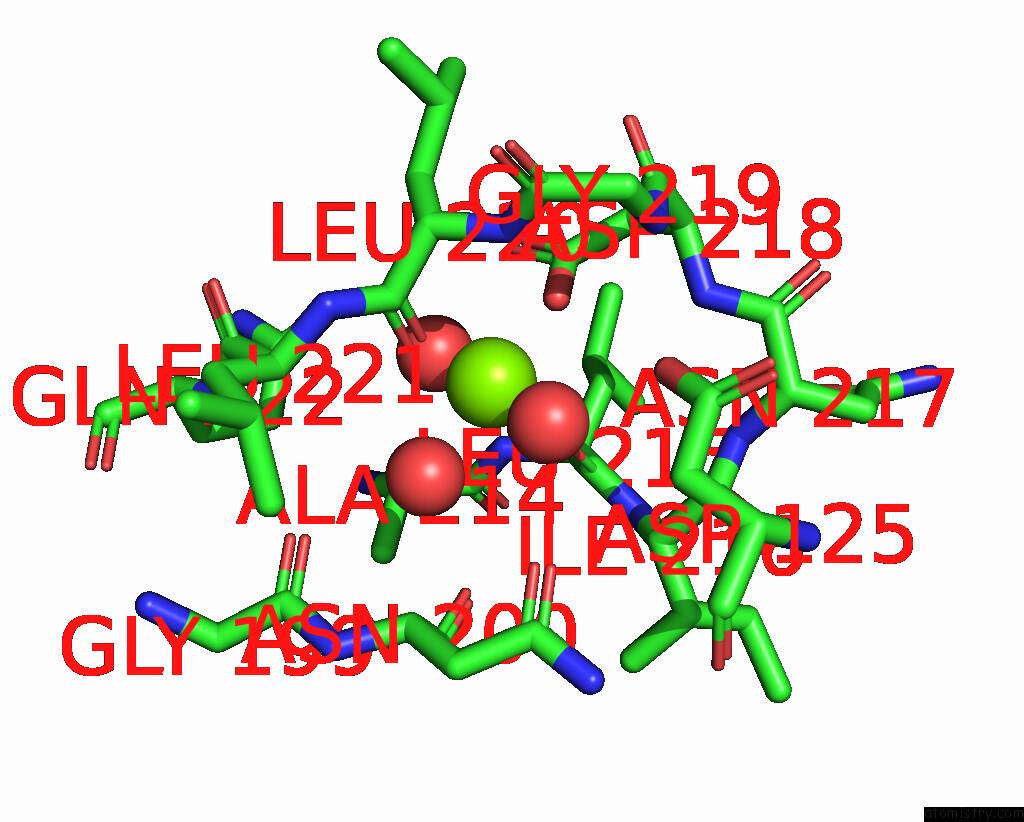
Mono view
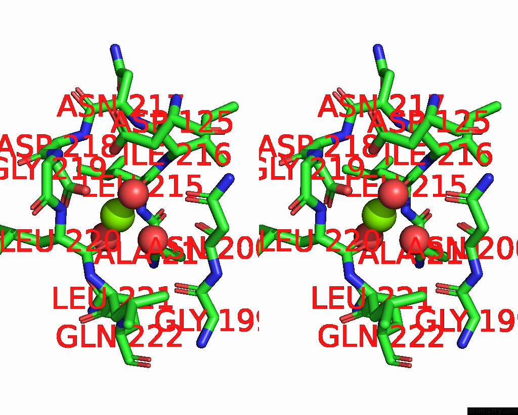
Stereo pair view
Mono view
Stereo pair view
A full contact list of Magnesium with other atoms in the Mg binding
site number 1 of Structure of An Escherichia Coli Lipid Kinase (Yegs) within 5.0Å range:
|
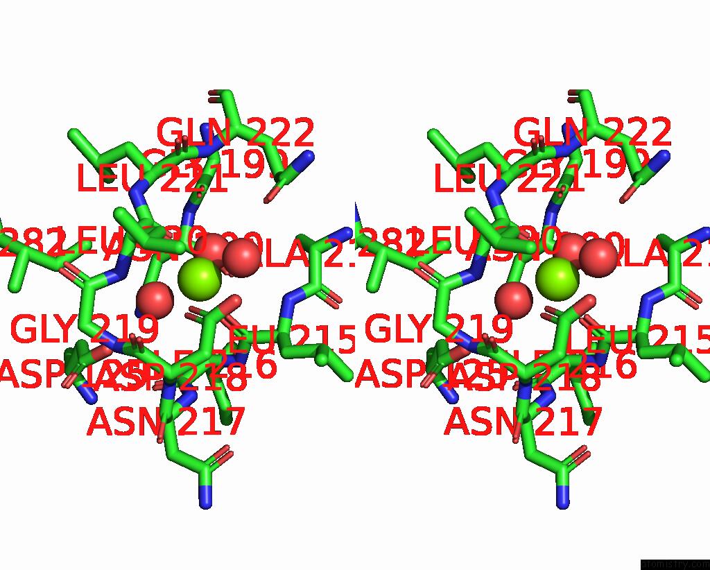
Magnesium binding site 2 out of 2 in 2bon
Go back to
Magnesium binding site 2 out
of 2 in the Structure of An Escherichia Coli Lipid Kinase (Yegs)

Mono view
Stereo pair view
Mono view
Stereo pair view
A full contact list of Magnesium with other atoms in the Mg binding
site number 2 of Structure of An Escherichia Coli Lipid Kinase (Yegs) within 5.0Å range:
|
Reference:
H.M.Bakali,
M.D.Herman,
K.A.Johnson,
A.A.Kelly,
A.Wieslander,
B.M.Hallberg,
P.Nordlund.
Crystal Structure of Yegs, A Homologue to the Mammalian Diacylglycerol Kinases, Reveals A Novel Regulatory Metal Binding Site. J.Biol.Chem. V. 282 19644 2007.
ISSN: ISSN 0021-9258
PubMed: 17351295
DOI: 10.1074/JBC.M604852200
Page generated: Tue Aug 13 21:57:56 2024
ISSN: ISSN 0021-9258
PubMed: 17351295
DOI: 10.1074/JBC.M604852200
Last articles
Zn in 9J0NZn in 9J0O
Zn in 9J0P
Zn in 9FJX
Zn in 9EKB
Zn in 9C0F
Zn in 9CAH
Zn in 9CH0
Zn in 9CH3
Zn in 9CH1