Magnesium »
PDB 2c42-2cic »
2c4r »
Magnesium in PDB 2c4r: Catalytic Domain of E. Coli Rnase E
Protein crystallography data
The structure of Catalytic Domain of E. Coli Rnase E, PDB code: 2c4r
was solved by
M.J.Marcaida,
A.J.Callaghan,
B.F.Luisi,
with X-Ray Crystallography technique. A brief refinement statistics is given in the table below:
| Resolution Low / High (Å) | 25.00 / 3.60 |
| Space group | P 62 2 2 |
| Cell size a, b, c (Å), α, β, γ (°) | 196.586, 196.586, 140.766, 90.00, 90.00, 120.00 |
| R / Rfree (%) | 31.9 / 34.7 |
Other elements in 2c4r:
The structure of Catalytic Domain of E. Coli Rnase E also contains other interesting chemical elements:
| Zinc | (Zn) | 1 atom |
Magnesium Binding Sites:
The binding sites of Magnesium atom in the Catalytic Domain of E. Coli Rnase E
(pdb code 2c4r). This binding sites where shown within
5.0 Angstroms radius around Magnesium atom.
In total 2 binding sites of Magnesium where determined in the Catalytic Domain of E. Coli Rnase E, PDB code: 2c4r:
Jump to Magnesium binding site number: 1; 2;
In total 2 binding sites of Magnesium where determined in the Catalytic Domain of E. Coli Rnase E, PDB code: 2c4r:
Jump to Magnesium binding site number: 1; 2;
Magnesium binding site 1 out of 2 in 2c4r
Go back to
Magnesium binding site 1 out
of 2 in the Catalytic Domain of E. Coli Rnase E
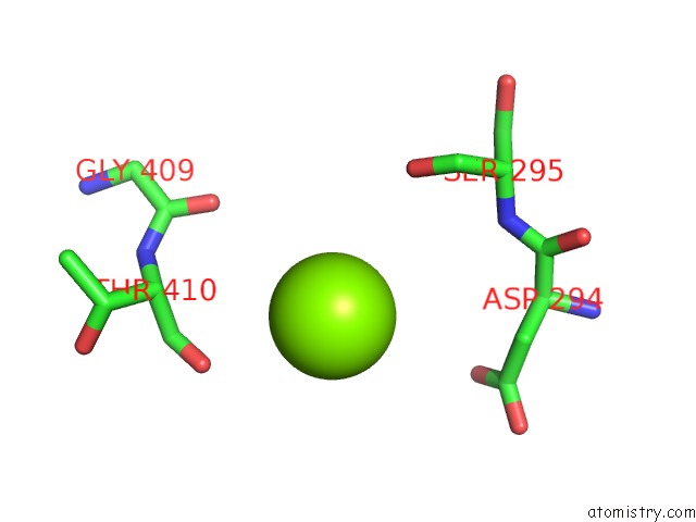
Mono view
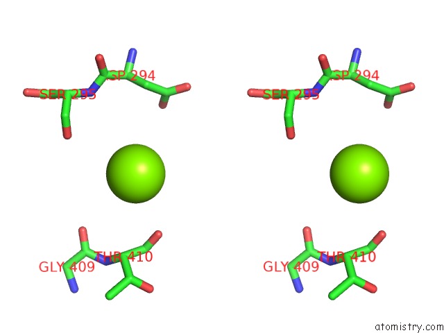
Stereo pair view
Mono view
Stereo pair view
A full contact list of Magnesium with other atoms in the Mg binding
site number 1 of Catalytic Domain of E. Coli Rnase E within 5.0Å range:
|
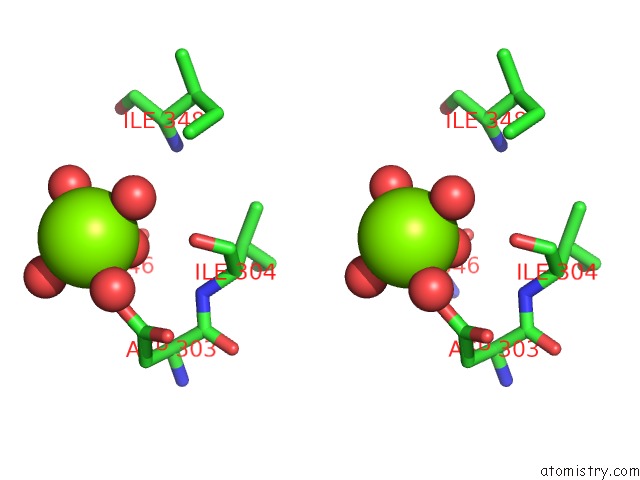
Magnesium binding site 2 out of 2 in 2c4r
Go back to
Magnesium binding site 2 out
of 2 in the Catalytic Domain of E. Coli Rnase E
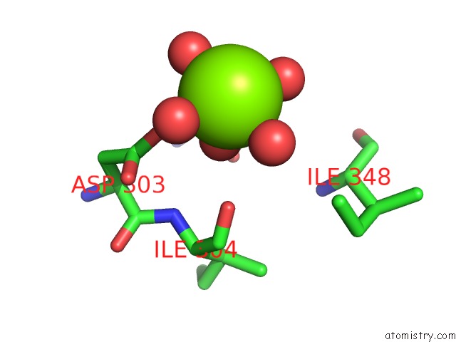
Mono view
Stereo pair view
Mono view
Stereo pair view
A full contact list of Magnesium with other atoms in the Mg binding
site number 2 of Catalytic Domain of E. Coli Rnase E within 5.0Å range:
|
Reference:
A.J.Callaghan,
M.J.Marcaida,
J.A.Stead,
K.J.Mcdowall,
W.G.Scott,
B.F.Luisi.
Structure of E. Coli Rnase E Catalytic Domain and Implications For Rna Processing and Turnover Nature V. 437 1187 2005.
ISSN: ISSN 0028-0836
PubMed: 16237448
DOI: 10.1038/NATURE04084
Page generated: Tue Aug 13 22:12:09 2024
ISSN: ISSN 0028-0836
PubMed: 16237448
DOI: 10.1038/NATURE04084
Last articles
Zn in 9J0NZn in 9J0O
Zn in 9J0P
Zn in 9FJX
Zn in 9EKB
Zn in 9C0F
Zn in 9CAH
Zn in 9CH0
Zn in 9CH3
Zn in 9CH1